Abstract
Macular pigment optical density (MPOD) quantifies the concentration of macular xanthophyll carotenoids—primarily lutein, zeaxanthin, and meso‑zeaxanthin—in the central retina.(1,2) These pigments are densely localized in the Henle fiber layer and photoreceptor axons of the fovea, where they filter short-wavelength blue light and exert antioxidant and anti-inflammatory effects.(1–3) Higher MPOD is associated with improved contrast sensitivity, glare disability, and photostress recovery, and observational evidence links low MPOD to increased risk of age-related macular degeneration (AMD) and possibly other retinal and optic nerve disorders.(2–5) MPOD is therefore considered a modifiable biomarker of retinal resilience and a potential intermediate endpoint for nutritional and therapeutic interventions.
Multiple techniques have been developed to measure MPOD in vivo, including heterochromatic flicker photometry (HFP), fundus reflectometry, fundus autofluorescence, resonance Raman spectroscopy, and more recently, artificial intelligence (AI)–based analysis of fundus images.(1,6–9) Each method has distinct advantages and limitations in terms of objectivity, repeatability, patient cooperation, and clinical feasibility.(1,6–9) Standard reference ranges for MPOD typically span 0–1 optical density units, with mean values around 0.30–0.35 in healthy adults.(10) Randomized trials show that supplementation with lutein and zeaxanthin increases MPOD in a dose-dependent manner and can improve aspects of visual performance, including contrast sensitivity and glare recovery, particularly in individuals with low baseline MPOD or early AMD.(3,4,11–13)
This review explains the anatomy and physiology of macular pigment, defines MPOD and how it is quantified, summarizes available measurement technologies, and synthesizes evidence linking MPOD to visual function and AMD risk. Emerging directions include AI-driven, non-invasive MPOD estimation from routine imaging, integration of MPOD with structural biomarkers such as optical coherence tomography (OCT)–derived retinal layer thickness, and evaluation of MPOD as an outcome measure in interventional trials.
Introduction
Macular pigment comprises the carotenoids lutein, zeaxanthin, and meso‑zeaxanthin concentrated in the central retina, giving the fovea its characteristic yellow appearance.(1,2) These pigments are obtained exclusively from the diet and accumulate selectively in the macula via complex transport mechanisms involving lipoproteins and binding proteins.(2,3) Functionally, macular pigment serves two key roles: optical filtering of high-energy visible (blue) light and biochemical quenching of reactive oxygen species.(2,3)
MPOD provides a quantitative measure of macular pigment at specified retinal eccentricities and has emerged as an important indicator of retinal health.(1,2) Lower MPOD has been associated with increased susceptibility to AMD and may correlate with structural and functional retinal changes even before overt disease becomes clinically apparent.(2,4,5,14) Conversely, macular carotenoid supplementation can increase MPOD and improve certain aspects of visual performance in healthy and diseased eyes.(3,4,11–13)
Understanding MPOD is therefore valuable both for basic research and for clinical practice, where it may inform nutritional counselling, risk stratification, and monitoring of intervention response.
Biology of Macular Pigment
Composition and Distribution
Macular pigment consists of three stereoisomeric xanthophyll carotenoids: lutein, zeaxanthin, and meso‑zeaxanthin.(2,3) Lutein predominates in the perifovea, while zeaxanthin and meso‑zeaxanthin are more concentrated in the central fovea.(2,3) These pigments are located primarily in the Henle fiber layer and inner plexiform layer, aligned along the paths of incident light to maximize filtering efficiency.(2,3)
Distribution typically shows a peaked profile centred on the fovea with exponential decline toward the parafovea. Inter-individual variability in MPOD and spatial distribution is substantial and influenced by diet, genetics, age, smoking status, and systemic factors.(2,3,10,14)
Optical and Antioxidant Functions
By absorbing blue light (400–500 nm), macular pigment reduces chromatic aberration and intraocular light scatter, thereby improving contrast sensitivity and reducing glare.(2,3,15) This optical filtering may be particularly relevant in bright or blue-light–rich environments, such as outdoor daylight or digital device use.
Biochemically, macular carotenoids quench singlet oxygen and other ROS, protect polyunsaturated fatty acids in photoreceptor outer segments from lipid peroxidation, and may modulate inflammatory pathways.(2,3,15,16) The combination of optical and antioxidant actions suggests a protective role against light-induced and oxidative damage to the macula.
Definition and Interpretation of MPOD
MPOD expresses the logarithmic ratio of macular pigment absorption at a given retinal location relative to a reference point with minimal pigment, typically in the perifovea.(1,6) Values are reported in optical density units, with higher values indicating greater pigment concentration. Population-based data suggest a typical range of 0–1.0, with mean MPOD around 0.35 and thresholds of approximately <0.21 (low), 0.21–0.44 (mid-range), and >0.44 (high), although exact cut-offs vary between studies and instruments.(10)
Higher MPOD is generally associated with:
- Better contrast sensitivity and reduced glare disability.
- Faster photostress recovery and improved dark adaptation.
- Lower prevalence or slower progression of AMD in some observational cohorts.(2–5,11–13,15–17)
However, MPOD is one of several interacting factors influencing retinal resilience; it should be interpreted alongside age, genetic risk, smoking history, diet, and structural imaging findings.
Methods for Measuring MPOD
A variety of in vivo techniques are used to assess MPOD, broadly categorized into psychophysical and objective optical or imaging methods.(1,6–9)
Heterochromatic Flicker Photometry (HFP)
HFP is the most widely used psychophysical technique and underpins several commercial instruments.(1,6,9) The method presents alternating blue (strongly absorbed by macular pigment) and green (minimally absorbed) light stimuli at specified retinal locations. The subject adjusts the relative intensity of the two wavelengths to minimize flicker perception; the difference in blue light intensity required at foveal versus perifoveal locations yields MPOD.
Advantages include relatively low cost and portability, but results depend on subject cooperation, fixation stability, and normal visual function, which may be challenging in elderly or diseased eyes.(1,6,9) Despite these limitations, HFP has good repeatability in experienced hands and has been used extensively in epidemiologic and interventional studies.(1,6,11–13)
Fundus Reflectometry
Fundus reflectometry measures spectral reflectance of the retina using specialized instruments and fits models of light absorption and scattering to derive macular pigment density.(1,6,7) It is an objective technique that does not rely on psychophysical responses but requires stable alignment, high-quality imaging, and complex calibration.(1,6,7) Variants include single-wavelength, dual-wavelength, and multi-spectral reflectometry.
Reflectometry can provide spatial maps of MP distribution but may be sensitive to ocular media opacity and requires careful correction for lens and macular reflections.(6,7)
Fundus Autofluorescence
Dual-wavelength fundus autofluorescence exploits the fact that macular pigment attenuates lipofuscin autofluorescence from the retinal pigment epithelium.(6,7) Images are acquired using two excitation wavelengths—one more strongly absorbed by macular pigment than the other—and the ratio of autofluorescence intensities is used to derive MPOD spatial maps.(6,7)
This method is objective and allows high-resolution topographic assessment but requires specialized imaging systems and careful standardization. It is increasingly used in research and some clinical settings and can be combined with OCT for multimodal assessment.(6,7)
Resonance Raman Spectroscopy
Resonance Raman spectroscopy measures characteristic vibrational signals from carotenoid molecules when excited with specific laser wavelengths.(6,8) It provides a direct, molecule-specific measure of carotenoid concentration with high specificity but requires laser-based instrumentation and is less widely available. Safety, cost, and technical complexity have limited its routine clinical use, though it remains a powerful research tool.(6,8)
Artificial Intelligence–Based Fundus Image Analysis
Recent work has proposed AI models that estimate MPOD from standard colour fundus photographs by segmenting macular regions and analysing colour channel information.(1,6,9,18) Deep learning algorithms can be trained against reference methods (for example, HFP or autofluorescence) to infer MPOD without dedicated hardware.(1,6,9,18) Narrative reviews suggest that AI-based approaches may offer scalable, non-invasive, and rapid MPOD screening, though rigorous validation against gold-standard techniques is ongoing.(1,6,9,18)
MPOD and Visual Function
Contrast Sensitivity and Glare
Systematic reviews and meta-analyses report positive correlations between MPOD and various measures of visual performance in healthy eyes.(2,15,19) Higher MPOD is linked to better contrast sensitivity at intermediate spatial frequencies and reduced disability glare, likely reflecting blue-light filtering and reduced intraocular scatter.(2,15,19)
In a meta-analysis of 22 publications, MPOD showed significant correlations with contrast sensitivity at specific eccentricities and spatial frequencies, and with photostress recovery time, particularly under low luminance conditions.(15) Interventional trials have demonstrated that increasing MPOD via lutein/zeaxanthin supplementation can improve contrast sensitivity and glare tolerance, especially in individuals with low baseline MPOD.(11–13,20)
Dark Adaptation and Photostress Recovery
Higher MPOD has been associated with faster recovery following photostress and better performance in low-luminance tasks, possibly through protection of photoreceptor and RPE function.(2,15,19) These parameters are clinically relevant in AMD and other macular disorders, where night vision difficulties are common.
Visual Function in AMD
In early AMD, reduced MPOD has been reported relative to age-matched controls, though findings vary by cohort and measurement method.(4,11–13) Randomized trials provide evidence that supplementing with lutein and zeaxanthin increases MPOD and can improve visual acuity, contrast sensitivity, and glare performance in patients with early or intermediate AMD.(11–13,20) These functional gains, while modest, may translate into meaningful benefits in daily visual tasks.
MPOD, AMD Risk, and Other Ocular Diseases
Associations with AMD
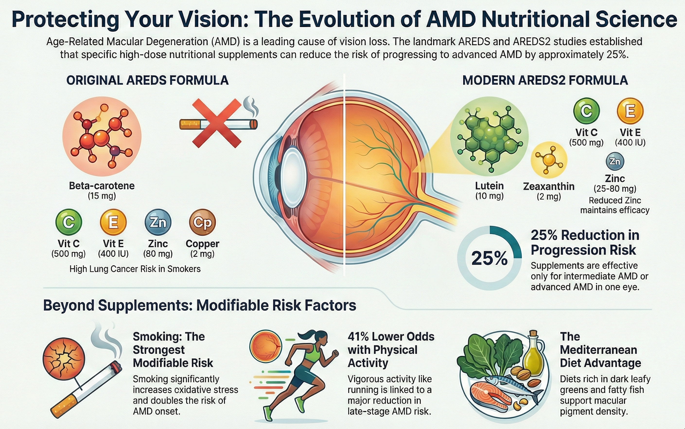
Observational studies suggest that lower MPOD is associated with higher prevalence of early AMD and possibly increased risk of progression, although causality is difficult to establish.(2,4,5,11,16) Low dietary intake of lutein and zeaxanthin correlates with both lower MPOD and higher AMD risk.(11,12,16) Some longitudinal cohorts indicate that individuals in the highest MPOD or dietary carotenoid tertiles have reduced risk of advanced AMD, aligning with findings from AREDS2 regarding lutein/zeaxanthin supplementation.(3–5,11–13,16)
Nonetheless, AMD is multifactorial, with strong contributions from age, genetics, smoking, and complement dysregulation. MPOD should therefore be viewed as one modifiable factor within a broader risk profile.
MPOD and Glaucoma
Emerging data suggest that MPOD may relate not only to macular but also to optic nerve health. In a large cohort from the CAREDS2 study, higher baseline MPOD was associated with greater central ganglion cell complex (GCC) and inner retinal layer thickness 15 years later, in both eyes with and without primary open-angle glaucoma.(5,21) These associations support the hypothesis that macular carotenoids may confer neuroprotective benefits beyond the macula, although interventional confirmation is required.
Other Conditions
Studies are exploring MPOD relationships with diabetic retinopathy, inherited retinal diseases, and visual performance in individuals with high digital device use, but data remain limited.(1,3,4,14,18) MPOD may also change following cataract surgery, given altered spectral input to the retina, which has implications for interpreting longitudinal measurements.(2,6)
Interventions to Modify MPOD
Nutritional Supplementation
Dietary and supplemental lutein and zeaxanthin reliably increase serum carotenoid levels and MPOD in most individuals, with a dose–response relationship and plateau effects at higher doses.(3,4,11–13,20) Randomized controlled trials show that daily doses of 10–20 mg lutein, alone or combined with 2–10 mg zeaxanthin, increase MPOD over 6–12 months and improve some visual function measures in early AMD and other populations.(11–13,20)
Individuals with low baseline MPOD or poor dietary intake often exhibit the largest relative gains.(11–13,20) Mesomeric carotenoids and formulations including meso‑zeaxanthin have also been shown to augment central MPOD, though large-scale AMD outcomes trials are limited.(12)
Lifestyle and Dietary Patterns
Dietary patterns rich in leafy green vegetables, coloured fruits, and egg yolks—major sources of lutein and zeaxanthin—are associated with higher MPOD and lower AMD prevalence.(11,12,16) Smoking, obesity, and low intake of carotenoid-rich foods are associated with lower MPOD.(2,11,16) Lifestyle counselling that encourages increased intake of macular carotenoids alongside AREDS2 supplementation may therefore be beneficial.
Emerging Research and Technological Directions
AI-Enhanced MPOD Measurement
Narrative reviews highlight the promise of AI-based fundus image analysis to estimate MPOD using standard non-mydriatic cameras, potentially enabling population-level screening and longitudinal monitoring in routine practice.(1,6,9,18) Deep learning models can segment macular regions and predict MPOD based on colour channel distributions, with early studies reporting encouraging agreement with reference methods.(1,6,9,18) Further validation across devices, ethnicities, and disease states is needed.
Structural Correlates of MPOD
Recent studies examine relationships between MPOD and OCT-derived retinal layer thicknesses, particularly in macular GCC and inner retinal layers.(5,21) Positive associations suggest that higher MPOD may be linked to preserved retinal structure over time, offering a potential bridge between nutritional biomarkers and neurodegenerative outcomes in diseases like glaucoma and AMD.(5,21)
MPOD as a Clinical Trial Endpoint
Given its modifiability and associations with visual function, MPOD is increasingly used as an outcome in trials of nutritional interventions and digital eye strain mitigation.(3,4,11–13,18,20) Standardization of measurement protocols and thresholds for clinically meaningful change will enhance its utility as a surrogate endpoint.
Conclusion
Macular pigment optical density provides a quantitative window into the concentration of lutein, zeaxanthin, and meso‑zeaxanthin in the central retina, reflecting both dietary intake and retinal carotenoid handling. Higher MPOD is linked to improved contrast sensitivity, reduced glare, and better photostress recovery, and lower MPOD has been associated with increased risk or severity of age-related macular degeneration and possibly other neurodegenerative eye diseases.(2–5,11–13,15–17,21)
Multiple psychophysical and objective methods allow MPOD measurement, and emerging AI-based techniques may broaden access to this biomarker in clinical practice.(1,6–9,18) Randomized trials show that macular carotenoid supplementation can increase MPOD and improve aspects of visual performance, particularly in individuals with low baseline levels or early AMD.(3,4,11–13,20) As research progresses, MPOD may serve not only as a marker of nutritional status but also as a component of integrated risk stratification and treatment monitoring frameworks for retinal disease.
This article is for educational purposes only and reflects current scientific literature at the time of writing.
References
- Zhang H, Li Y, Wang X, et al. Macular pigment optical density and measurement technology based on artificial intelligence: a narrative review. Int J Ophthalmol. 2025;18(6):40534793. https://pubmed.ncbi.nlm.nih.gov/40534793/[pubmed.ncbi.nlm.nih]
- Bernstein PS, Delori FC, Richer S, et al. The value of measurement of macular carotenoid pigment optical densities and distributions in age-related macular degeneration and other retinal disorders. Vision Res. 2010;50(7):716–728. https://pmc.ncbi.nlm.nih.gov/articles/PMC8169741/[pmc.ncbi.nlm.nih]
- Ma L, Dou HL, Wu YQ, et al. Lutein and zeaxanthin and their roles in age-related macular degeneration. J Ophthalmol. 2013;2013:120864. https://pmc.ncbi.nlm.nih.gov/articles/PMC6774801/[pmc.ncbi.nlm.nih]
- Beatty S, Chakravarthy U, Nolan JM, et al. Secondary outcomes in a clinical trial of macular pigment supplementation in patients with age-related macular degeneration. Invest Ophthalmol Vis Sci. 2013;54(7):4561–4570. https://pmc.ncbi.nlm.nih.gov/articles/PMC6774801/[pmc.ncbi.nlm.nih]
- Loewen R, Myers CE, Rogers JD, et al. Macular pigment optical density as a measurable modifiable biomarker of retinal health. Clin Ophthalmol. 2024;18:11478551. https://pmc.ncbi.nlm.nih.gov/articles/PMC11478551/[pmc.ncbi.nlm.nih]
- Howells O, Eperjesi F, Bartlett HE. Measuring macular pigment optical density in vivo: a review of techniques. Graefes Arch Clin Exp Ophthalmol. 2011;249(3):315–347. https://pmc.ncbi.nlm.nih.gov/articles/PMC8169741/[pmc.ncbi.nlm.nih]
- van der Veen RLP, Berendschot TTJM, Makridaki M, et al. An optical method to assess the macular pigment density. Invest Ophthalmol Vis Sci. 2014;55(1):446–454. https://lo.um.es/arvo-abstract/an-optical-method-to-assess-the-macular-pigment-density/[lo.um]
- Bernstein PS, Gellermann W. Measurement of carotenoid concentrations in human retina and skin. Methods Enzymol. 2002;319:315–330. https://pmc.ncbi.nlm.nih.gov/articles/PMC8169741/[pmc.ncbi.nlm.nih]
- Li J, Wang Y, Zhang L, et al. Macular pigment optical density and measurement technology based on artificial intelligence: opportunities and challenges. Ophthalmol Ther. 2025;14(3):ed478cb28bd94a4aafd954d6529c1f47. https://paperity.org/p/366256560[paperity]
- Wikipedia contributors. Macular pigment optical density. Wikipedia. 2025. https://en.wikipedia.org/wiki/Macular_pigment_optical_density[en.wikipedia]
- Huang YM, Dou HL, Huang FF, et al. Effect of lutein and zeaxanthin on macular pigment and visual function in patients with early age-related macular degeneration. Ophthalmology. 2012;119(11):2290–2297. https://www.sciencedirect.com/science/article/abs/pii/S0161642012005453[sciencedirect]
- Nolan JM, Akkali MC, Loughman J, et al. Macular pigment response to supplementation with meso‑zeaxanthin, lutein, and zeaxanthin in persons with low macular pigment: the Meso‑zeaxanthin Ocular Supplementation Trial. Invest Ophthalmol Vis Sci. 2012;53(9):6114–6123. https://pmc.ncbi.nlm.nih.gov/articles/PMC6774801/[pmc.ncbi.nlm.nih]
- Liu R, Wang T, Zhang B, et al. Macular pigment response to lutein, zeaxanthin, and meso‑zeaxanthin supplementation in glaucomatous eyes: a randomized controlled trial. J Glaucoma. 2021;30(7):595–603. https://pubmed.ncbi.nlm.nih.gov/36247822/[pubmed.ncbi.nlm.nih]
- Renzi LM, Hammond BR Jr. The relation between the macular carotenoids, lutein and zeaxanthin, and visual performance. Nutrients. 2010;2(12):1200–1212. https://pmc.ncbi.nlm.nih.gov/articles/PMC8169741/[pmc.ncbi.nlm.nih]
- Loughman J, Akkali MC, Beatty S, et al. The association between macular pigment optical density and visual performance: a systematic review and meta-analysis. Ophthalmic Physiol Opt. 2020;40(5):423–446. https://pmc.ncbi.nlm.nih.gov/articles/PMC8169741/[pmc.ncbi.nlm.nih]
- Seddon JM, Ajani UA, Sperduto RD, et al. Dietary carotenoids and vitamins A, C, and E and advanced age-related macular degeneration. JAMA. 1994;272(18):1413–1420. https://pubmed.ncbi.nlm.nih.gov/7933422[pmc.ncbi.nlm.nih]
- Carotenoids in Age-Related Eye Disease Study 2 (CAREDS2) Research Group. Association of macular pigment optical density with retinal layer thicknesses in eyes with and without manifest primary open-angle glaucoma. BMJ Open Ophthalmol. 2023;8(1):e001331. https://bmjophth.bmj.com/content/8/1/e001331[bmjophth.bmj]
- Wang X, Zhang H, Gao Y, et al. Artificial intelligence-based measurement of macular pigment optical density from fundus photographs. Transl Vis Sci Technol. 2025;14(2):12. https://doaj.org/article/ed478cb28bd94a4aafd954d6529c1f47[doaj]
- Hammond BR Jr, Wooten BR, Snodderly DM. Individual variations in the spatial profile of human macular pigment. J Opt Soc Am A. 1997;14(6):1187–1196. https://pmc.ncbi.nlm.nih.gov/articles/PMC8169741/[pmc.ncbi.nlm.nih]
- Stringham JM, Hammond BR Jr. The glare hypothesis of macular pigment function. Optom Vis Sci. 2007;84(9):859–864. https://pmc.ncbi.nlm.nih.gov/articles/PMC8169741/[pmc.ncbi.nlm.nih]
Leave a comment
This site is protected by hCaptcha and the hCaptcha Privacy Policy and Terms of Service apply.