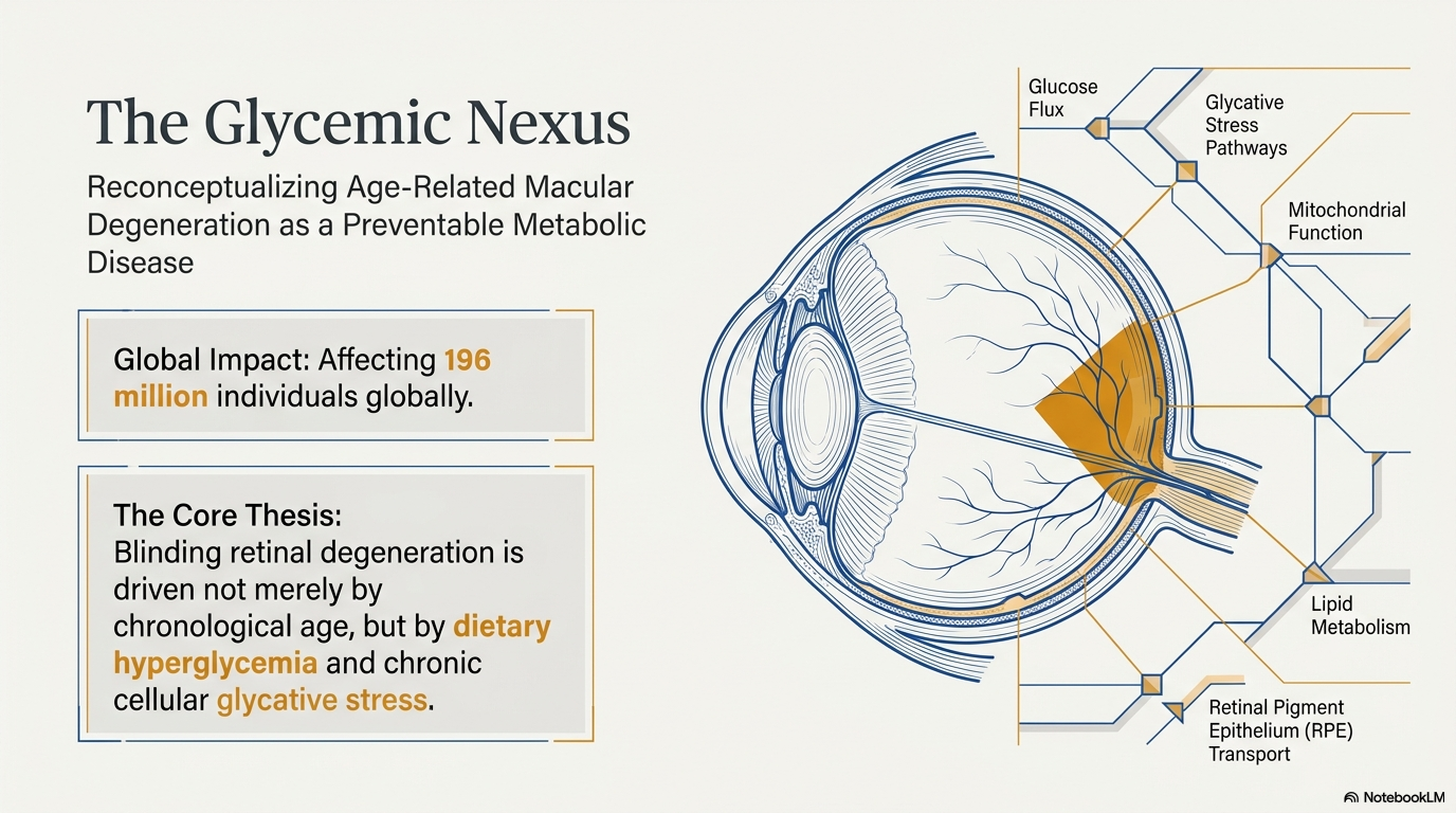
The Glycemic Nexus: Impact of Dietary Hyperglycemia and Advanced Glycation End-Products on Age-Related Macular Degeneration
Age-related macular degeneration (AMD) represents the leading cause of irreversible blindness and visual impairment in individuals over the age of 50 in industrialized nations, currently affecting approximately 196 million people globally. As life expectancy continues to rise and global populations age, the prevalence of this multifactorial neurodegenerative disease is projected to escalate dramatically, placing an unprecedented burden on healthcare infrastructure and individual quality of life. AMD is characterized by the progressive degeneration of the retinal pigment epithelium (RPE), Bruch's membrane, and the overlying photoreceptor cells in the macula—the central region of the retina responsible for high-acuity vision. While genetic susceptibility and chronological aging remain primary risk factors, a burgeoning body of research has identified a critical metabolic driver: the "Glycemic Nexus". This framework posits that dietary hyperglycemia—driven by the rapid absorption of sugars from high-glycemic index (GI) diets—triggers chronic glycative stress, leading to the accumulation of cytotoxic Advanced Glycation End-Products (AGEs) and the subsequent failure of cellular proteostasis in the retina.

Pathophysiology of Glycative Stress: The Molecular Mechanisms of Retinal Injury
The fundamental molecular process underpinning the Glycemic Nexus is the non-enzymatic reaction between reducing sugars and biological macromolecules, a process traditionally known as the Maillard reaction. In the context of the human retina, this process is not merely a marker of aging but a primary pathogenic mechanism.
The Biochemistry of Advanced Glycation End-Products (AGEs)
Glycation begins when a reducing sugar, such as glucose, or a reactive dicarbonyl intermediate like methylglyoxal (MGO), reacts with the amino groups of proteins, lipids, or nucleic acids. This initial interaction forms an unstable Schiff base, which undergoes a chemical rearrangement over several weeks to form a more stable Amadori product.These products eventually transition into irreversible, cross-linked structures known as Advanced Glycation End-Products (AGEs).
MGO is a particularly potent precursor in this pathway, rapidly modifying proteins to form MG-H1 conjugates.Unlike enzymatic reactions, glycation is largely driven by the concentration of the reactants—primarily the spikes in blood glucose that occur following the consumption of high-GI carbohydrates. These AGEs are inherently cytotoxic, forming insoluble protein aggregates and disrupting the biochemical properties of the extracellular matrix (ECM). In the eye, AGEs accumulate in the cornea, lens, vitreous, and significantly within the RPE and Bruch's membrane, where they alter structural proteins and promote a state of chronic metabolic dysfunction.

Retinal Pigment Epithelium Vulnerability and the Phagocytic Burden
The RPE occupies a unique and demanding physiological niche. It is a monolayer of post-mitotic cells that must function for the duration of a human life without the possibility of "diluting" cellular damage through division. Each RPE cell is tasked with the daily phagocytosis and digestion of the distal 10% of approximately 30 photoreceptor outer segments (POSs) to maintain the visual cycle. This relentless metabolic activity creates an immense proteolytic burden.
Under conditions of dietary hyperglycemia, the high influx of glucose accelerates the formation of AGEs within the RPE. The accumulation of these glycated proteins triggers a "vicious cycle". First, the glycation of POS proteins renders them resistant to normal enzymatic degradation. Second, the AGEs themselves directly inhibit the cell's own cleanup crews: the ubiquitin-proteasome system (UPS) and the autophagy-lysosomal pathway.

Failure of the Cellular Cleanup Machinery
The UPS is responsible for the degradation of short-lived or damaged soluble proteins, while autophagy handles larger aggregates and organelles. Glycative stress disrupts these systems through multiple avenues:
- Substrate Modification: Glycation transforms proteins into forms that are inaccessible to proteases, such as the alpha A162 protein, which becomes resistant to UPS-dependent degradation upon glycation.
- Inactivation of Protective Machinery: Reactive dicarbonyls like MGO damage the proteolytic enzymes and ubiquitin themselves, reducing overall protein turnover rates.
- Proteolytic Overload: The accumulation of dark, insoluble, and cross-linked AGEs (often referred to as brunescent proteins) eventually overwhelms the lysosomal capacity, leading to the build-up of lipofuscin—an undegradable fluorescent waste product that is a hallmark of retinal aging.
When these systems fail, the RPE can no longer maintain its homeostatic balance, leading to the formation of extracellular drusen—deposits of lipids and proteins between the RPE and Bruch's membrane—which further impede the flow of nutrients and oxygen to the photoreceptors.

Cellular Senescence and the Senescence-Associated Secretory Phenotype (SASP)
As the RPE accumulates damage that it can neither repair nor dilute, many of these cells enter a state of cellular senescence. Senescence is characterized by a permanent growth arrest accompanied by profound phenotypic changes, including cellular flattening, hypertrophy, and resistance to apoptosis.
The "Zombie" Cell State in AMD
Senescent RPE cells are often described as "zombie" cells because they are no longer functional in their primary roles but remain metabolically active. They exhibit a Senescence-Associated Secretory Phenotype (SASP), through which they secrete a potent cocktail of pro-inflammatory cytokines and growth factors.
|
SASP Component Category |
Specific Factors Identified |
Impact on Retinal Environment |
|
Pro-inflammatory Cytokines |
IL-1\alpha, IL-1\beta, IL-6, IL-8, TNF-\alpha, IFN-\gamma |
Drives chronic low-grade inflammation ("inflammaging") |
|
Growth Factors |
VEGF-A |
Promotes abnormal blood vessel growth (neovascularization) in wet AMD |
|
Immune Regulators |
IL-10, various chemokines |
Recruits and activates microglial cells and macrophages |
These secretions exert a "bystander effect," where the senescent cells poison their healthy neighbors, leading to the spread of geographic atrophy (dry AMD) or the initiation of neovascularization (wet AMD). Molecular triggers of this senescence include oxidative stress, DNA damage, and the accumulation of A2E—a toxic byproduct of the visual cycle that can be exacerbated by blue light exposure.

Dietary Glycemic Index (GI) and AMD: Epidemiological and Clinical Foundations
The Glycemic Index is a physiological measure that ranks carbohydrate-containing foods based on how quickly they raise blood glucose levels after ingestion. Foods with a high GI (e.g., white bread, sweetened cereals, refined starches) produce rapid glucose spikes, whereas low-GI foods (e.g., whole grains, legumes, non-starchy vegetables) release glucose gradually into the bloodstream.

Insights from the AREDS Cohort
The Age-Related Eye Disease Study (AREDS) provided some of the most compelling evidence linking dietary carbohydrate quality to AMD risk. In an analysis of 4,099 nondiabetic participants, researchers categorized individuals by their dietary glycemic index (dGI).
|
Dietary GI Quintile |
Comparison to Lowest Quintile |
Outcome for AMD Features |
|
Highest Quintile |
OR: 1.42 (1.09 - 1.84) |
Significantly increased risk of large drusen |
|
Highest Quintile |
OR: 1.78 (0.81 - 3.90) |
Suggestive increased risk of geographic atrophy |
|
Highest Quintile |
OR: 1.41 (0.95 - 2.08) |
Suggestive increased risk of neovascularization |
The study concluded that if the participants had simply consumed a diet with a GI below the sex-specific median (≥ 77.9 for women; ≥ 79.3 for men), 20% of prevalent cases of AMD in that population would have been eliminated.Furthermore, longitudinal data indicated that individuals with high dGI intake had an 8% to 17% higher risk of disease progression over five years compared to those with low dGI intake.
Animal Corroboration and the "Rescue" Effect
Animal models have successfully replicated these findings, providing mechanistic proof of the Glycemic Nexus. C57BL/6 mice fed high-GI diets for prolonged periods developed multiple AMD-like features, including RPE vacuolation, loss of basal infoldings, and the accumulation of AGEs. Remarkably, when aged mice were switched from a high-GI to a low-GI diet, the progression of retinal damage was arrested, and some features were partially reversed.This suggests that the metabolic environment of the retina is dynamic and that dietary modification can provide a "metabolic rescue" even in later life.
The Gut-Eye Axis: Microbiome Modulation by Diet
Modern research has expanded the Glycemic Nexus to include the gut microbiome, revealing that the impact of high-GI foods is mediated by intestinal health. High-glycemic diets alter the microbial composition of the gut, promoting dysbiosis that triggers systemic inflammation.
 Microbial Profiles and AMD Susceptibility
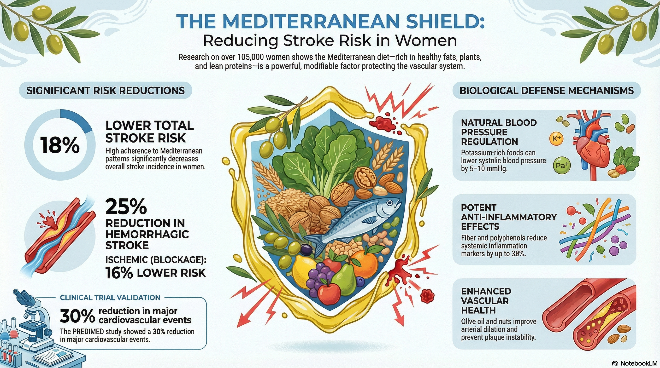
Studies have identified specific bacterial populations associated with AMD risk. Diets high in refined sugars promote an abundance of the order Clostridiales, which is linked to increased AMD susceptibility. Conversely, low-GI diets, such as the Mediterranean diet, promote the growth of Bacteroidales, which are associated with protection against retinal degeneration.
The Mediterranean diet—rich in fish, vegetables, olive oil, and whole grains—acts as a multi-modal intervention by:
- Strengthening the Intestinal Barrier: Reducing the translocation of inflammatory markers into the systemic circulation.
- Producing Beneficial Metabolites: Increasing the production of short-chain fatty acids (SCFAs) like butyrate, which have potent anti-inflammatory effects.
- Elevating Protective Signaling: Low-GI diets increase circulating levels of serotonin and other protective metabolites that modulate the retinal immune response.

Comprehensive Nutritional Interventions and Supplementation
The nutritional management of AMD has advanced significantly since the original AREDS trial. Current standard-of-care recommendations focus on the AREDS2 formulation while incorporating emerging evidence on polyphenols and specific dietary patterns.
The AREDS2 Formulation
The landmark AREDS2 trial established the optimal blend of vitamins and minerals for slowing the progression of intermediate-to-advanced AMD. A critical change from the original formula was the replacement of Beta-carotene with Lutein and Zeaxanthin to eliminate the increased risk of lung cancer in smokers.
|
Supplement Component |
Daily Dosage (AREDS2) |
Role in Retinal Health |
|
Vitamin C |
500 mg |
Primary water-soluble antioxidant |
|
Vitamin E |
400 IU |
Protects cell membranes from lipid peroxidation |
|
Zinc (Zinc Oxide) |
80 mg |
Essential for enzyme function in the visual cycle |
|
Copper (Cupric Oxide) |
2 mg |
Prevents copper-deficiency anemia caused by high zinc |
|
Lutein |
10 mg |
Forms macular pigment; filters blue light |
|
Zeaxanthin |
2 mg |
Central macular pigment; antioxidant |
While effective for those with existing disease, AREDS2 supplements are not intended for prevention in healthy individuals, for whom dietary sources are preferred.
The Synergistic Dietary Compound Score
Research indicates that the combined effect of a low-GI diet and the AREDS2 nutrients is greater than the sum of its parts. Scientists developed a "Dietary Compound Score" that aggregates the intake of Lutein/Zeaxanthin, Zinc, Vitamins C and E, Omega-3 fatty acids (DHA/EPA), and a low-GI profile. High scores on this composite measure were robustly associated with the greatest reduction in the risk of both prevalent drusen and advanced AMD.
Saffron: A "Golden" Neuroprotectant
Saffron (Crocus sativus) and its active constituents, crocin and crocetin, have emerged as potent adjunct therapies.Clinical evidence suggests that daily supplementation with 20–50 mg of saffron for 3 to 12 months significantly improves visual acuity and retinal function as measured by electroretinograms (ERG).
Saffron's mechanisms include:
- Anti-angiogenic Activity: Counteracting the VEGF cascade to inhibit abnormal vessel growth.
- Neuroprotection: Protecting retinal cells from light-induced damage by inhibiting apoptosis pathways.
- Antioxidant Support: Depleting free radicals during periods of acute oxidative stress.
Curcumin and the CBNS Revolution
Curcumin, the primary polyphenol in turmeric, has recently demonstrated a remarkable association with reduced AMD risk. A massive retrospective cohort study published in 2024 utilized electronic health records from over 1.8 million patients and found that the use of Curcuma-Based Nutritional Supplements (CBNS) was associated with significantly lower rates of all forms of AMD.
|
Outcome for Patients Aged 50+ |
Risk Ratio (CBNS vs. Control) |
Statistical Significance |
|
Developing any AMD |
0.23 |
P < 0.001 |
|
Advanced Dry AMD (GA) |
0.11 |
P < 0.001 |
|
Exudative (Wet) AMD |
0.28 |
P < 0.001 |
|
Legal Blindness |
0.46 |
P < 0.001 |
|
Need for Anti-VEGF Injections |
0.15 |
P < 0.001 |
For patients with early nonexudative AMD, the introduction of CBNS was associated with a 42% reduction in the risk of progressing to late-stage disease (RR: 0.58).
Environmental and Lifestyle Modifications
Beyond nutrition, the Glycemic Nexus interacts with physical activity and environmental light exposure to determine retinal longevity.
Physical Activity and Vascular Stability
Vigorous physical activity has been shown to exert a protective effect against both early and late AMD. A meta-analysis of longitudinal studies involving 14,000 patients revealed that high levels of activity (e.g., swimming, running, tennis) significantly reduced the risk of early AMD development.
Specifically, running distance has a dose-response relationship with AMD risk. For every additional kilometer run per day, the relative risk for AMD decreased by 10%. Individuals running more than 4 km per day had a 42% to 54% lower risk than sedentary individuals, independent of diet or smoking status. This is likely due to exercise's role in improving systemic vascular health, lowering blood pressure, and reducing oxidative stress.
Protecting the Eye from Phototoxicity
The retina is highly sensitive to the blue and ultraviolet (UV) portions of the light spectrum. UV radiation is divided into three segments:
- UVC (<286 nm): Filtered by the ozone layer.
- UVB (286–320 nm): Causes sunburn and is mostly absorbed by the cornea.
- UVA (320–400 nm): The most concerning for retinal health, as it is transmitted to the lens and contributes to cumulative oxidative damage.
Blue light (400–500 nm) is particularly damaging because it reacts with chemicals in the retina to produce free radicals.Melanin, the body's natural sunscreen, absorbs this harmful light, but its concentration in the RPE decreases by approximately 50% by age 65, increasing vulnerability to AMD.

Practical Protection Recommendations:
- UV400 Sunglasses: Ensure sunglasses block 100% of UVA and UVB rays up to 400 nm.
- Blue-Blocking Tints: Lenses with yellow, gray, or brown tints effectively block blue light and reduce glare without significantly darkening the field of vision.
- Wide-Brimmed Hats: A simple hat can reduce direct UV exposure significantly.
- Digital Hygiene: Use software like f.lux or built-in "night shift" modes on devices to reduce blue light intensity during screen time.

Future Horizons: Senolytics and Metabolic Modulators
The treatment of AMD is moving beyond passive supplementation toward active metabolic intervention.
Senolytic Therapies
Given the role of RPE senescence in disease progression, researchers are investigating "senolytics"—drugs that selectively eliminate these dysfunctional cells. A promising candidate is Mini Cry, a 20-mer peptide derived from \alpha B-crystallin, which has been shown to decrease the number of senescent RPE cells and suppress the secretion of SASP components like VEGF.
Metformin and SGLT2 Inhibitors
Metformin, widely used for diabetes, is being studied for its ability to decrease the progression of geographic atrophy in non-diabetic patients (the METforMIN trial). By improving mitochondrial function and activating cellular cleanup pathways, metformin may mimic some of the protective effects of a low-GI diet. Additionally, SGLT2 inhibitors (e.g., empagliflozin) have shown a significant association with lower risks of AMD and dry AMD in diabetic populations compared to other medications, highlighting the importance of glucose control in preserving vision.

Strategic Guidance: The Eye Health Food Pyramid
Synthesizing the vast body of nutritional research into a practical framework leads to the development of the Eye Health Food Pyramid. This model prioritizes foods that stabilize the Glycemic Nexus and provide essential macular nutrients.
|
Frequency |
Dietary Recommendation |
Rationale |
|
Daily |
3 servings of Low-GI Whole Grains (Oats, Basmati rice, Quinoa) |
Maintains stable blood glucose; prevents glycative stress |
|
Daily |
5 servings of Dark Leafy Greens & Colorful Fruits (Kale, Spinach, Berries) |
High in Lutein, Zeaxanthin, and Vitamin C |
|
Daily |
2-3 cups of Green or Black Tea |
Catechins and caffeine provide neuroprotection and anti-inflammatory support |
|
Weekly |
2-4 servings of Oily Fish (Salmon, Sardines, Mackerel) |
Rich in Omega-3 (DHA/EPA) for retinal membrane integrity |
|
Weekly |
2-3 handfuls of Nuts and Seeds (Almonds, Walnuts) |
Sources of Vitamin E and healthy fats |
|
Regularly |
Anti-inflammatory Spices (Saffron, Turmeric/Curcumin) |
Potent radical scavengers and VEGF inhibitors |
|
Limit |
High-GI Foods (White bread, Sugary sodas, Fried snacks) |
Accelerates AGE formation and RPE damage |
FAQ: Scientific Perspectives on Common Concerns
Q: Can dietary changes really "reverse" AMD? A: While clinical neovascularization (wet AMD) requires immediate medical treatment, evidence from animal models suggests that switching to a low-GI diet can halt the progression of early-to-intermediate lesions and may partially restore the metabolic integrity of the RPE. In humans, lowering dGI is one of the most effective ways to slow progression.
Q: Are carrots the best food for vision? A: This is a partial myth. Carrots provide Vitamin A, which is essential for general vision, but dark leafy greens (kale, spinach) are much more effective for preventing AMD because they contain the specific carotenoids (lutein/zeaxanthin) that form the protective macular pigment.
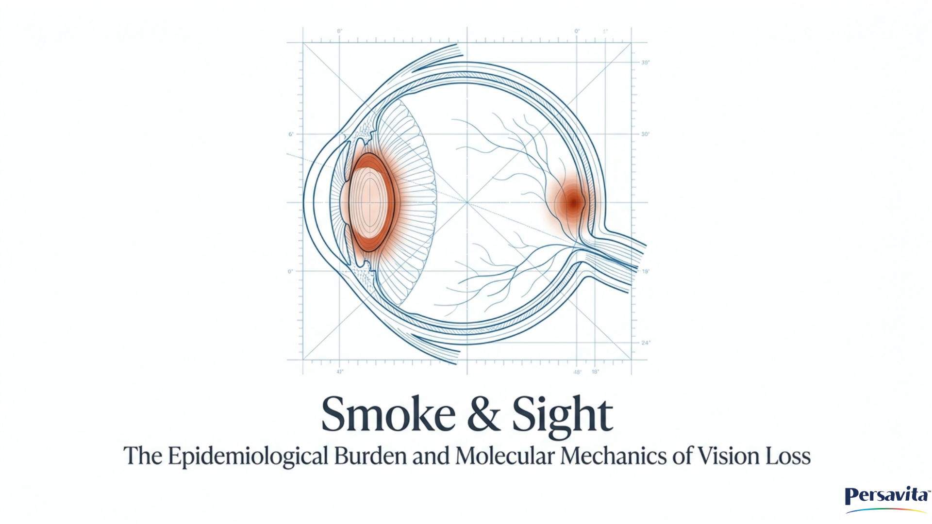
Q: Does sugar directly cause blindness? A: Indirectly, yes. Excessive intake of high-GI carbohydrates causes chronic hyperglycemia, which generates "glycotoxins" called AGEs. These compounds damage the RPE cells that keep the retina alive. A high-GI diet is considered a risk factor comparable to smoking for AMD development.
Q: Should I take eye vitamins if I have a family history but no symptoms? A: The AREDS2 formulation is scientifically proven to help only those who have already reached the intermediate or advanced stages of AMD. For those at risk but without symptoms, the focus should be on a Mediterranean-style, low-GI diet and the lifestyle habits mentioned above.
Q: Does screen use cause AMD? A: Screen use causes temporary eye strain, dryness, and fatigue, but it has not been definitively proven to cause AMD. However, the blue light emitted by screens may accelerate retinal aging in individuals who already have other risk factors, so using blue-light filters is a prudent preventive measure.
Q: What is a "senolytic," and is it a treatment for AMD? A: Senolytics are a new class of drugs designed to clear out "zombie" (senescent) RPE cells that are no longer functioning but are secreting inflammatory factors. While not yet standard treatment, medications like Mini Cry and others are currently in various stages of research to determine if they can prevent the spread of geographic atrophy.
Conclusion: A Paradigm Shift in Retinal Care
The Glycemic Nexus represents a foundational shift in how we understand and manage Age-Related Macular Degeneration. By recognizing that the retina is a mirror of our systemic metabolic health, we can implement strategies that go beyond simple supplementation. The evidence underscores a powerful truth: the choices we make regarding carbohydrate quality, environmental protection, and physical activity are the most potent tools we have to preserve vision for a lifetime. As the population ages, the widespread adoption of these evidence-based metabolic interventions offers a tangible path toward reducing the global incidence of blindness and empowering individuals to maintain their visual independence well into their later years.
|
Intervention Category |
Specific Component |
Dosage or Frequency |
Primary Role in Retinal Health |
Key Evidence or Outcome |
Target Population |
Source |
|
Supplement |
Lutein |
10 mg |
Forms macular pigment; filters blue light |
Optimal blend for slowing progression in intermediate-to-advanced AMD cases. |
Individuals with intermediate-to-advanced AMD |
|
|
Supplement |
Zeaxanthin |
2 mg |
Central macular pigment; antioxidant |
Replacement for Beta-carotene in AREDS2 to eliminate lung cancer risk in smokers. |
Individuals with intermediate-to-advanced AMD |
|
|
Supplement |
Zinc (Zinc Oxide) |
80 mg |
Essential for enzyme function in the visual cycle |
Part of the AREDS2 formulation found effective in slowing disease progression. |
Individuals with intermediate-to-advanced AMD |
|
|
Supplement |
Saffron (Crocus sativus) |
20–50 mg daily |
Anti-angiogenic; neuroprotection; antioxidant support |
Significant improvements in visual acuity and retinal function (ERG) over 3-12 months. |
Individuals with existing AMD |
|
|
Supplement |
Curcuma-Based Nutritional Supplements (CBNS) |
Not in source |
Potent radical scavengers and VEGF inhibitors |
42% reduction in risk of progressing to late-stage disease; lower rates of legal blindness. |
Individuals aged 50+; patients with early nonexudative AMD |
|
|
Dietary |
Dark Leafy Greens (Kale, Spinach) |
5 servings daily |
High in Lutein and Zeaxanthin to form protective macular pigment |
More effective than carrots for preventing AMD due to specific carotenoid content. |
Healthy individuals for prevention |
|
|
Dietary |
Oily Fish (Salmon, Sardines, Mackerel) |
2–4 servings weekly |
Rich in Omega-3 (DHA/EPA) for retinal membrane integrity |
High Dietary Compound Scores (including Omega-3) associated with greatest risk reduction. |
General population; those at risk of AMD |
|
|
Dietary |
Low-GI Whole Grains |
3 servings daily |
Maintains stable blood glucose; prevents glycative stress |
AREDS cohort data suggests 20% of cases could be eliminated by consuming low-GI diets. |
Nondiabetic individuals; healthy individuals for prevention |
|
|
Lifestyle |
Vigorous Physical Activity (e.g., Running) |
> 4 km per day |
Improving systemic vascular health and reducing oxidative stress |
42% to 54% lower risk of AMD compared to sedentary individuals. |
General population; individuals at risk of early AMD |
|
|
Lifestyle |
UV400 Sunglasses & Blue-Blocking Tints |
Regularly during light exposure |
Filters UVA/UVB and high-energy blue light to prevent free radical production |
Protects RPE from phototoxicity, which is critical as melanin concentration decreases with age. |
Individuals aged 65+; individuals with existing risk factors |
|
Works cited
- Healthy Dietary Patterns and Risk of Major Eye Diseases: Evidence ..., accessed April 15, 2026, https://pmc.ncbi.nlm.nih.gov/articles/PMC12954465/
- The effect of caffeine consumption on age-related macular degeneration: a systematic review and meta-analysis | Request PDF - ResearchGate, accessed April 15, 2026, https://www.researchgate.net/publication/391430041_The_effect_of_caffeine_consumption_on_age-related_macular_degeneration_a_systematic_review_and_meta-analysis
- Glycative stress as a cause of macular degeneration - PubMed - NIH, accessed April 15, 2026, https://pubmed.ncbi.nlm.nih.gov/?term=%22Avila-Portillo%20N%22[Author]
- Glycative stress as a cause of macular degeneration - PMC - NIH, accessed April 15, 2026, https://pmc.ncbi.nlm.nih.gov/articles/PMC11699537/
- Targeting Lysosomal Dysfunction and Oxidative Stress in Age-Related Macular Degeneration - PMC, accessed April 15, 2026, https://pmc.ncbi.nlm.nih.gov/articles/PMC12108406/
- Debunking Common Myths About Age-Related Macular Degeneration, accessed April 15, 2026, https://www.crmd.net/debunking-common-myths-about-age-related-macular-degeneration/
- Advanced Glycation End Products in Disease Development and Potential Interventions, accessed April 15, 2026, https://pmc.ncbi.nlm.nih.gov/articles/PMC12024296/
- Mechanisms of RPE senescence and potential role of αB crystallin ..., accessed April 15, 2026, https://pmc.ncbi.nlm.nih.gov/articles/PMC8923947/
- Autophagy in Age-Related Macular Degeneration: A Regulatory Mechanism of Oxidative Stress - PMC, accessed April 15, 2026, https://pmc.ncbi.nlm.nih.gov/articles/PMC7429811/
- Natural history of age-related retinal lesions that precede AMD in mice fed high or low glycemic index diets - PubMed, accessed April 15, 2026, https://pubmed.ncbi.nlm.nih.gov/22205601/
- Full article: Autophagy and heterophagy dysregulation leads to retinal pigment epithelium dysfunction and development of age-related macular degeneration - Taylor & Francis, accessed April 15, 2026, https://www.tandfonline.com/doi/full/10.4161/auto.24546
- The gut-eye axis in age-related macular degeneration: from microbial dysbiosis to targeted intervention strategies - PMC, accessed April 15, 2026, https://pmc.ncbi.nlm.nih.gov/articles/PMC12864145/
- Age-Associated Decline in Autophagy Pathways in Retinal Pigment Epithelium and Protective Effects of Topical Trehalose in Light-induced Outer Retinal Degeneration in Mice | bioRxiv, accessed April 15, 2026, https://www.biorxiv.org/content/10.1101/2025.01.14.632938v1.full-text
- Does senescence play a role in age-related macular degeneration? - PMC, accessed April 15, 2026, https://pmc.ncbi.nlm.nih.gov/articles/PMC10032649/
- Aging-US: RPE cell senescence in age-related macular degeneration, accessed April 15, 2026, https://www.aging-us.com/news-room/rpe-cell-senescence-in-age-related-macular-degeneration
- Can Blue Light and Sun Exposure Impact Macular Degeneration? - Eye Hub Optometry, accessed April 15, 2026, https://www.eyehubhtx.com/blog/can-blue-light-and-sun-exposure-impact-macular-degeneration.html
- Association between dietary glycemic index and age-related ..., accessed April 15, 2026, https://pubmed.ncbi.nlm.nih.gov/17616779/
- Dietary carbohydrate and the progression of age-related macular degeneration: a prospective study from the Age-Related Eye Disease Study - PubMed, accessed April 15, 2026, https://pubmed.ncbi.nlm.nih.gov/17921404/
- Nutritional Supplements for Age-Related Macular Degeneration - PMC - NIH, accessed April 15, 2026, https://pmc.ncbi.nlm.nih.gov/articles/PMC7015456/
- New Study Confirms the Efficacy of AREDS2 Eye Vitamin Supplement for Slowing Age-Related Macular Degeneration - BrightFocus, accessed April 15, 2026, https://www.brightfocus.org/resource/new-study-confirms-the-efficacy-of-areds2-eye-vitamin-supplement-for-slowing-age-related-macular-degeneration/
- Nutrition for eye health and macular disease, accessed April 15, 2026, https://www.macularsociety.org/support/daily-life/practical-guides/healthy-living/nutrition/
- NCT00345176 | Age-Related Eye Disease Study 2 (AREDS2) | ClinicalTrials.gov, accessed April 15, 2026, https://clinicaltrials.gov/study/NCT00345176
- Myths and Facts about AMD - AMD | Patient & Caregiver - Blindness Prevention, accessed April 15, 2026, https://patient-amd.vision-relief.com/myths-and-facts-about-amd/
- Dietary Compound Score and Risk of Age-Related Macular ..., accessed April 15, 2026, https://pmc.ncbi.nlm.nih.gov/articles/PMC3753024/
- Crocus sativus (saffron) and age-related macular degeneration - PMC, accessed April 15, 2026, https://pmc.ncbi.nlm.nih.gov/articles/PMC11537240/
- Short-term Outcomes of Saffron Supplementation in Patients with Age-related Macular Degeneration: A Double-blind, Placebo-controlled, Randomized Trial - PMC, accessed April 15, 2026, https://pmc.ncbi.nlm.nih.gov/articles/PMC5342880/
- Pharmacological Effects of Saffron and its Constituents in Ocular Disorders from in vitro Studies to Clinical Trials: A Systematic Review - PMC, accessed April 15, 2026, https://pmc.ncbi.nlm.nih.gov/articles/PMC8033960/
- Curcumin in Retinal Diseases: A Comprehensive Review from Bench to Bedside - PMC, accessed April 15, 2026, https://pmc.ncbi.nlm.nih.gov/articles/PMC8998602/
- Curcuma-based nutritional supplements and reduced risk of AMD - Modern Retina, accessed April 15, 2026, https://www.modernretina.com/view/curcuma-based-nutritional-supplements-and-reduced-risk-of-amd
- Curcuma-Based Nutritional Supplements and Risk of Age-Related Macular Degeneration, accessed April 15, 2026, https://pubmed.ncbi.nlm.nih.gov/39446346/
- Curcuma-Based Nutritional Supplements and Risk of Age-Related Macular Degeneration, accessed April 15, 2026, https://pmc.ncbi.nlm.nih.gov/articles/PMC11581721/
- Physical Activity and Age-related Macular Degeneration: A Systematic Literature Review and Meta-analysis | Request PDF - ResearchGate, accessed April 15, 2026, https://www.researchgate.net/publication/317185673_Physical_Activity_and_Age-related_Macular_Degeneration_A_Systematic_Literature_Review_and_Meta-analysis
- Physical Activity and AMD Risk - American Academy of Ophthalmology, accessed April 15, 2026, https://www.aao.org/eyenet/article/physical-activity-and-amd-risk
- High Levels of Physical Activity Protect Against AMD - American Academy of Ophthalmology, accessed April 15, 2026, https://www.aao.org/eyenet/article/physical-activity-protects-against-amd
- Prospective Study of Incident Age-Related Macular Degeneration in Relation to Vigorous Physical Activity during a 7-Year Follow-up - PMC, accessed April 15, 2026, https://pmc.ncbi.nlm.nih.gov/articles/PMC4090325/
- Exercise Intervention for Age-Related Macular Degeneration · Recruiting Participants for Clinical Trial 2026 - withpower.com, accessed April 15, 2026, https://www.withpower.com/trial/exercise-intervention-for-age-related-macular-degeneration-f9d59
- Ultra-violet and Blue Light Worsen Macular Degeneration - AMDF, accessed April 15, 2026, https://www.macular.org/about-macular-degeneration/what-is-macular-degeneration/risk-factors/ultra-violet-and-blue-light
- Ultra-violet and Blue Light Aggravate Macular Degeneration - Mills Vision Care, accessed April 15, 2026, https://www.millsvisioncare.com/ultra-violet-and-blue-light-aggravate-macular-degeneration/
- Protect Your Eyes from Bright and Blue Lights - BrightFocus, accessed April 15, 2026, https://www.brightfocus.org/resource/protect-your-eyes-from-bright-and-blue-lights/
- Protecting your eyes from glare and UV - Macular Society, accessed April 15, 2026, https://www.macularsociety.org/support/daily-life/practical-guides/healthy-living/protecting-your-eyes/
- Targeting senescent retinal pigment epithelial cells facilitates retinal regeneration in mouse models of age-related macular degeneration - PMC, accessed April 15, 2026, https://pmc.ncbi.nlm.nih.gov/articles/PMC8602547/
- Study Details | NCT02684578 | Metformin for the Minimization of Geographic Atrophy Progression in Patients With AMD | ClinicalTrials.gov, accessed April 15, 2026, https://clinicaltrials.gov/study/NCT02684578
- Use of SGLT2 Inhibitors Versus DPP-4 Inhibitors and Age-Related Macular Degeneration in Patients WithType 2 Diabetes: A Multinational Cohort Study - PMC, accessed April 15, 2026, https://pmc.ncbi.nlm.nih.gov/articles/PMC12020956/
- Debunking Macular Degeneration Myths: What You Need to Know - Rosen Optometry, accessed April 15, 2026, https://www.rosenoptometry.com/debunking-macular-degeneration-myths-what-you-need-to-know/
- Caffeine intake and late dry age-related macular degeneration: tea's protective role—insights from NHANES and Mendelian randomization - ResearchGate, accessed April 15, 2026, https://www.researchgate.net/publication/400833988_Caffeine_intake_and_late_dry_age-related_macular_degeneration_tea's_protective_role-insights_from_NHANES_and_Mendelian_randomization
- Green Tea Benefits for Seniors - Antioxidants for the Eyes, Brain, and Body, accessed April 15, 2026, https://naturaleyecare.com/blog/green-tea-benefits-for-seniors-antioxidants-for-the-eyes-brain-and-body/
- Features That Distinguish Age-Related Macular Degeneration from Aging - PMC - NIH, accessed April 15, 2026, https://pmc.ncbi.nlm.nih.gov/articles/PMC11975485/
Leave a comment
This site is protected by hCaptcha and the hCaptcha Privacy Policy and Terms of Service apply.