
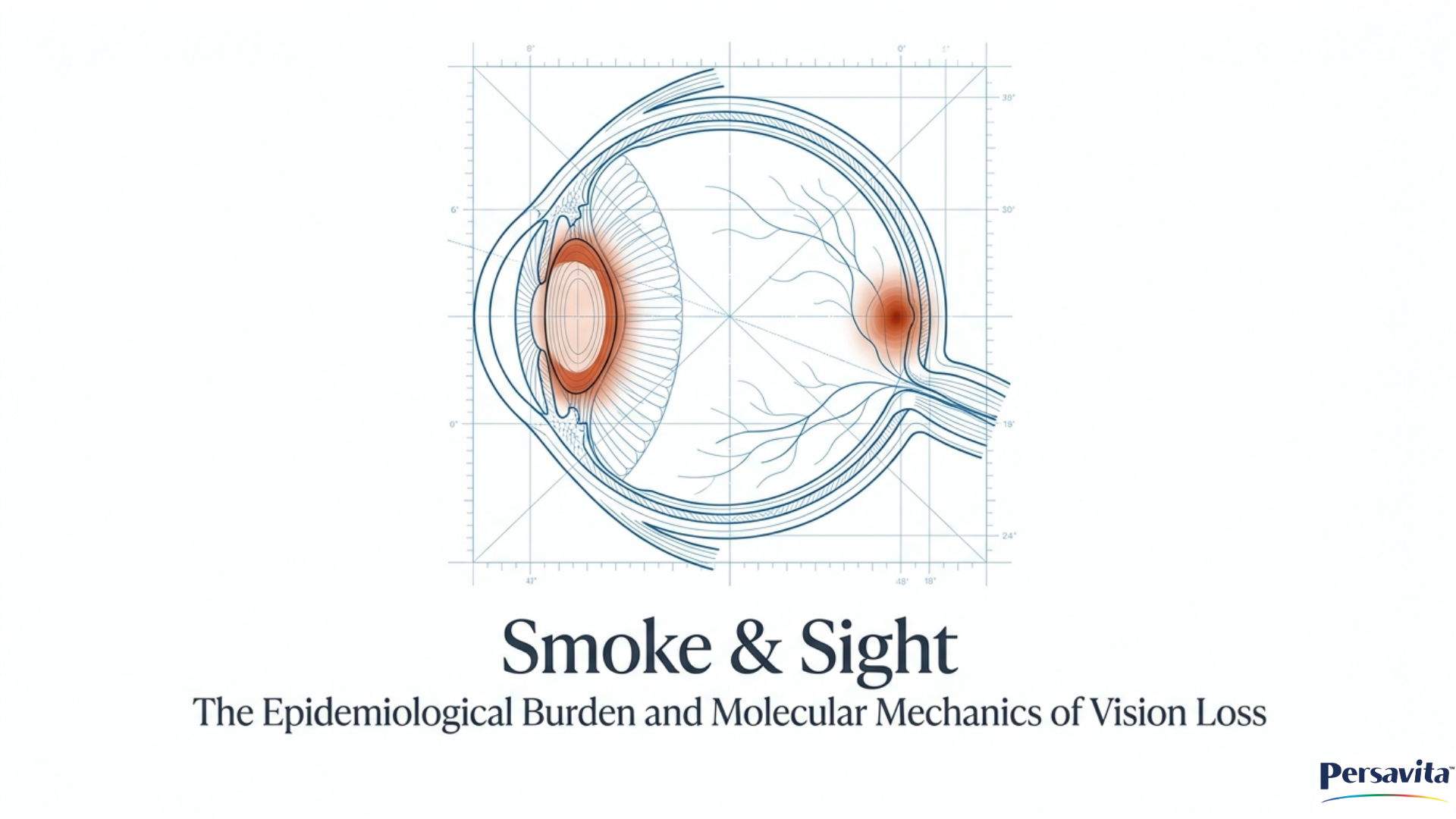
Could High Uric Acid Affect Your Eye Health?
New Research Explores a Potential Link to Age-Related Macular Degeneration
Recent clinical research findings robustly establish a significant association between gout—and its underlying metabolic driver, hyperuricemia—and an increased risk of developing Age-Related Macular Degeneration (AMD).
Large-Scale Population Studies Clinical understanding of the link between gout and AMD has been heavily shaped by two major observational studies that examined massive patient datasets:
- The 2018 Medicare Cohort Study: An analysis of 5% of Medicare claims data, representing over 1.6 million U.S. adults aged 65 and older, identified gout as an independent risk factor for incident AMD. The incidence rate of AMD was 20.1 per 1,000 person-years for people with gout, compared to 11.7 for those without. After controlling for potential confounders, a diagnosis of gout was associated with a 39% increased hazard of developing AMD.
- The 2025 Multi-Center Database Study: A more recent retrospective study analyzed data from over 16,000 propensity-score matched patient pairs across 68 U.S. healthcare organizations. The researchers found that gout significantly increases the risk of developing and progressing to severe forms of AMD. Specifically, over a 5-year period, patients with gout were 2.73 times more likely to develop dry AMD, 2.64 times more likely to develop advanced dry AMD (geographic atrophy), and 2.48 times more likely to develop wet AMD. Furthermore, gout patients were significantly more likely to suffer macular hemorrhages and require anti-VEGF injections. Among patients who already had early AMD, the presence of gout drastically worsened outcomes, increasing the risk of progression to advanced dry AMD by more than 10-fold and to wet AMD by over 3-fold.
- Clinical Case Reports In addition to epidemiological data, direct clinical observations have noted specific, unusual retinal complications in patients with gout. For instance, a case report of a 62-year-old man with chronically uncontrolled gout detailed the discovery of highly refractile, crystal-like lesions in his macula. These lesions, which did not stain with fluorescein and were associated with areas of geographic atrophy, were believed to be direct uric acid deposits located at the level of the retinal pigment epithelium (RPE).
The Larger Context: Shared Mechanisms of Inflammation and Oxidative Stress These clinical findings highlight that gout is not merely a localized joint disease, but a systemic condition with serious implications for eye health. Researchers hypothesize that the connection between gout and AMD is rooted in shared pathological mechanisms:
- Chronic Inflammation: Both conditions are heavily driven by chronic, systemic inflammation. In gout, elevated uric acid leads to monosodium urate (MSU) crystals that trigger the NLRP3 inflammasome, an inflammatory pathway that releases cytokines like IL-1β. Similarly, in AMD, drusen deposits trigger the exact same NLRP3 inflammasome activation within the retinal pigment epithelium (RPE) cells, contributing to their death and photoreceptor dysfunction.
- The Uric Acid Paradox and Oxidative Stress: While uric acid acts as an antioxidant in the bloodstream by scavenging free radicals, it can become a harmful pro-oxidant inside cells. Inside retinal tissues, elevated uric acid levels can promote oxidative stress, lipid peroxidation, and cellular damage, leading to long-term retinal degeneration.
- Proposed "Hyperuricemic AMD" Pathways: Researchers have proposed the concept of "hyperuricemic AMD," suggesting the disease could develop via two concurrent routes. The "crystal-induced pathway" suggests urate crystals deposit directly in the choroid capillary–Bruch's membrane–RPE complex causing local inflammation and drusen formation. The "non-crystal-induced pathway" suggests hyperuricemia triggers the renin-angiotensin system (RAS), which actively degrades the retinal complex and induces abnormal blood vessel growth (neovascularization) seen in wet AMD.
- Together, these clinical findings and mechanistic theories suggest that managing metabolic health and uric acid levels may be an important, previously under-recognized component of preserving long-term retinal health and slowing AMD progression. Future research is focused on whether urate-lowering therapies could definitively improve ocular outcomes for these patients
Could High Uric Acid Affect Your Eye Health?
When people think about uric acid, they usually think about gout, a painful condition caused by the buildup of uric acid crystals in the joints. However, emerging research suggests that elevated uric acid levels may affect more than just the joints. Scientists are now investigating whether hyperuricemia (high uric acid levels in the blood) could also play a role in age-related macular degeneration (AMD), one of the leading causes of vision loss among older adults.

Understanding AMD
Age-related macular degeneration affects the macula, the central part of the retina responsible for sharp, detailed vision. AMD can make it difficult to read, recognize faces, drive, and perform many everyday activities.
Although age remains the strongest risk factor, researchers have identified several additional factors that may influence AMD development and progression, including smoking, genetics, cardiovascular health, obesity, and chronic inflammation.

What Is Hyperuricemia?
Hyperuricemia occurs when uric acid levels become elevated in the bloodstream. While uric acid is a natural byproduct of metabolism, excessive amounts may contribute to inflammation and oxidative stress throughout the body.
Persistently elevated uric acid levels can lead to gout, but researchers are increasingly interested in how hyperuricemia may affect other tissues, including the eyes.

The Emerging Connection Between Uric Acid and AMD
A growing number of studies have reported an association between gout, elevated uric acid levels, and AMD.
A recent review published in The FASEB Journal highlighted evidence suggesting that hyperuricemia may contribute to retinal inflammation, oxidative stress, retinal pigment epithelium (RPE) dysfunction, and abnormal blood vessel growth, all processes implicated in AMD development.
Researchers have proposed that high uric acid levels may activate inflammatory pathways, including the NLRP3 inflammasome, which has already been linked to AMD. Elevated uric acid may also increase oxidative stress within retinal cells and promote damage to structures that support retinal health.

Large Study Finds Increased AMD Risk Among Patients With Gout
In 2025, researchers analyzed data from more than 16,000 patients with gout and compared them with a matched control group.
The study found that people with gout were:
• 2.7 times more likely to develop dry AMD
• 2.6 times more likely to develop advanced dry AMD (geographic atrophy)
• 2.5 times more likely to develop wet AMD
• More likely to experience retinal bleeding
• More likely to require anti-VEGF injections used to treat wet AMD
Among individuals who already had early AMD, gout was also associated with a significantly higher risk of progression to advanced disease.
While these findings do not prove that gout causes AMD, they strengthen the evidence that metabolic health and chronic inflammation may influence long-term retinal health.

Why Might Uric Acid Affect the Retina?
Interestingly, uric acid appears to have a dual nature.
In the bloodstream, uric acid can act as an antioxidant and help neutralize harmful free radicals.
Inside cells, however, elevated uric acid may have the opposite effect. Research suggests it may promote oxidative stress, inflammatory signaling, and cellular damage. This imbalance could potentially contribute to retinal degeneration over time.
Researchers are continuing to investigate exactly how these mechanisms interact within retinal tissues.

What This Means for Eye Health
Although more research is needed, these findings reinforce an important message:
Eye health is closely connected to overall health.
Maintaining healthy metabolic function may benefit not only the heart, joints, and blood vessels but also the retina.
Practical steps that support long-term eye health include:
• Regular comprehensive eye examinations
• Following medical advice to manage gout and elevated uric acid levels
• Maintaining a healthy body weight
• Eating a balanced diet rich in fruits and vegetables
• Avoiding smoking
• Staying physically active
• Using evidence-based nutritional support when appropriate

Looking Ahead
Scientists are continuing to investigate whether lowering uric acid levels could help protect retinal health or slow AMD progression. While definitive answers are not yet available, the growing body of evidence highlights the importance of metabolic health as part of a comprehensive approach to healthy aging and vision preservation.
As our understanding of AMD continues to evolve, factors once considered unrelated to eye health, such as uric acid metabolism, may prove to be important pieces of the puzzle.


Frequently asked Questions (FAQs):
Can urate crystals be directly seen in the macula?
Yes, while historically very rare, urate crystals can be directly observed in the macula,.
A specific clinical case report documented this phenomenon in a 62-year-old man with a long history of severe, chronically uncontrolled gout,. Upon presenting with blurred vision, an examination revealed highly refractile, crystal-like lesions in his macula,.
Key characteristics of these visible macular deposits included:
- Location: The crystals were subretinal, located specifically at the level of the retinal pigment epithelium (RPE), and were predominantly clustered at the termination of retinal arterioles,.
- Appearance: They presented as small, yellow, highly refractile lesions that notably did not stain with fluorescein dye during angiography,.
- Association with Atrophy: The crystal deposits were directly associated with larger areas of geographic atrophy (advanced dry AMD) in the patient's macula,.
- Asymmetric Presentation: The crystal deposits were primarily unilateral (affecting only his left eye), which researchers noted correlates with the way gout typically presents asymmetrically in the body's joints,.
-
Prior to this specific case, while gout had been linked to other ocular complications and an increased risk of macular degeneration, there had not been any documented reports of direct urate crystal deposits in the retina. The documentation of these lesions, which researchers concluded were consistent with uric acid deposits, underscores the importance of dilated retinal exams for patients with gout who experience visual complaints.
What is the 'uric acid paradox' and how does it affect the eyes?
The "uric acid paradox" refers to the contradictory, dual nature of uric acid depending on whether it is located inside or outside of a cell.
In the Bloodstream (Antioxidant) Outside of cells, such as in the bloodstream, circulating uric acid functions as a highly beneficial antioxidant. It helps neutralize harmful free radicals, acts as an iron chelator to curb iron-mediated oxidative stress, and counters strong oxidants like peroxynitrite. Through these mechanisms, it provides a protective role that includes shielding against lipid oxidation within blood cell membranes.
Inside Cells (Pro-oxidant) Conversely, when elevated uric acid enters the intracellular environment, it has the exact opposite effect and acts as a harmful pro-oxidant. The intracellular degradation of uric acid generates free radicals. Furthermore, uric acid can directly elevate the activity of NADPH oxidase, an enzyme that significantly amplifies the production of superoxide free radicals inside the cell.
How It Affects the Eyes In the context of eye health, this paradoxical shift from antioxidant to pro-oxidant can be highly destructive. Inside retinal tissues, the pro-oxidant behavior of elevated uric acid promotes oxidative stress, lipid peroxidation, and severe cellular damage.
This intracellular stress is believed to trigger a cascade of negative effects in the eye, including:
- Inflammatory Signaling: It can activate inflammatory pathways, such as the NLRP3 inflammasome, which has been directly linked to Age-Related Macular Degeneration (AMD).
- RPE Dysfunction: It damages the retinal pigment epithelium (RPE), the layer of cells that nourishes and supports the retina.
- Abnormal Blood Vessel Growth: It may contribute to the pathological growth of new, fragile blood vessels in the retina.
Ultimately, the cellular damage and chronic inflammation caused by intracellular uric acid can contribute to long-term retinal degeneration, which researchers increasingly suspect is a mechanism driving the development and progression of AMD.
What is the crystal-induced pathway for macular degeneration?
The "crystal-induced pathway" is a hypothesized mechanism explaining how high uric acid levels (hyperuricemia) may drive the development and progression of Age-Related Macular Degeneration (AMD).
This pathway is theorized to closely resemble the way gouty arthritis damages joints, but occurring within the delicate tissues of the eye. The process involves the following steps:
- Crystal Deposition: Uric acid (UA) or monosodium urate (MSU) crystals physically form and deposit within the choroid capillary–Bruch's membrane–retinal pigment epithelium (RPE) complex.
- Inflammatory Activation: The physical presence of these crystals triggers a chronic inflammatory response, specifically by activating the NLRP3 inflammasome pathway.
- Cellular Damage (Pyroptosis): The activation of this inflammasome leads to the generation of reactive oxygen species (ROS) and induces pyroptosis, a highly inflammatory form of programmed cell death that destroys the RPE cells.
- AMD Hallmarks: This cascade of chronic inflammation and cellular destruction ultimately results in the classic clinical signs of AMD, including drusen formation, the accumulation of cellular debris (lipofuscin), geographic atrophy (advanced dry AMD), and choroidal neovascularization (the abnormal blood vessel growth characteristic of wet AMD).
This pathway is one of two proposed routes for "hyperuricemic AMD," with the other being a non-crystal-induced pathway driven by the activation of the renin-angiotensin system (RAS).
What is the non-crystal-induced pathway involving the RAS?
The non-crystal-induced pathway describes a mechanism by which high uric acid levels (hyperuricemia) can damage the retina without the physical formation and deposition of urate crystals. Instead, this pathway is driven by the abnormal activation of the renin-angiotensin system (RAS).
Here is how researchers hypothesize this biochemical pathway unfolds:
- RAS Activation and Angiotensin II Surge: Hyperuricemia triggers the RAS, causing a significant increase in the expression and levels of angiotensin II (Ang II) in the eyes.
- Cellular Signaling Cascade: The excess Ang II binds to its receptor (AT1R), which sets off a chain reaction of signaling inside the retinal pigment epithelium (RPE) cells. This includes the phosphorylation and activation of specific signaling proteins, such as ERK and p38 MAPK.
- Release of Destructive Enzymes and Cytokines: This signaling cascade causes the RPE and retinal ganglion cells to produce elevated levels of destructive enzymes, particularly matrix metalloproteinases like MMP-2 and MMP-14, as well as basigin (BSG). Furthermore, the RAS activation triggers the production of inflammatory cytokines, including TNF-α, IL-6, and IL-1β.
- Tissue Degradation and Neovascularization: The release of these MMP enzymes actively breaks down and degrades the delicate choroid capillary–Bruch's membrane–RPE complex that supports the retina. Simultaneously, Ang II upregulates VEGFR-2 and potentiates the activity of vascular endothelial growth factor (VEGF). This combination of structural degradation and VEGF stimulation induces abnormal, leaky blood vessel growth (choroidal neovascularization), which is the primary hallmark of wet AMD.
Ultimately, this RAS-driven cascade leads to severe damage of the RPE cell junctions, triggers cell death (apoptosis), and drives the progression of macular degeneration.
How do matrix metalloproteinases contribute to retinal tissue degradation?
Matrix metalloproteinases (MMPs) contribute to retinal tissue degradation by physically breaking down the delicate support structures of the retina, a process primarily driven by the "non-crystal-induced pathway" associated with high uric acid levels.
Here is how these enzymes specifically drive this degradation:
- Triggered by the Renin-Angiotensin System (RAS): Elevated uric acid (hyperuricemia) abnormally activates the RAS, causing a surge in angiotensin II (Ang II) levels in the eye.
- Enzyme Activation and Production: The excess Ang II signals the retinal pigment epithelium (RPE) cells and retinal ganglion cells to ramp up the production and activation of specific destructive enzymes, most notably MMP-14 (alongside a protein called basigin) and MMP-2.
- Structural Breakdown: Once activated, these matrix metalloproteinases go to work on the eye's architecture. They actively degrade the choroid capillary–Bruch's membrane–RPE complex. This complex is crucial for nourishing and supporting the retina.
- Perpetuating Retinal Damage: By breaking down this vital support complex, MMPs disrupt the physical junctions and interactions between cells, which perpetuates retinal damage, causes RPE cell death, and ultimately facilitates the progression of age-related macular degeneration (AMD).
What is the relationship between hyperuricemia and wet AMD?
Epidemiological data and mechanistic research demonstrate a strong, direct relationship between hyperuricemia (the metabolic driver of gout) and a significantly increased risk of developing wet AMD.
Significantly Increased Clinical Risk Large-scale database studies indicate that individuals with gout and hyperuricemia are approximately 2.5 times more likely to develop wet AMD over a 5-year period compared to those without the condition. The danger is even more pronounced for patients who already have early-stage AMD; the presence of gout increases their risk of progressing to the wet form of the disease by more than 3-fold (a 3.32 relative risk). Because of this rapid progression to neovascularization, hyperuricemic patients are significantly more likely to suffer from macular hemorrhages and require anti-VEGF injections, which are the standard treatment for wet AMD.
Biological Mechanisms Driving Wet AMD Mechanistically, elevated serum uric acid levels have been directly linked to the onset of choroidal neovascularization, the pathological and fragile blood vessel growth that clinically defines wet AMD.
As noted in our previous discussion of the "non-crystal-induced pathway," hyperuricemia can damage the retina by abnormally activating the renin-angiotensin system (RAS). This biochemical cascade specifically drives the development of wet AMD through several angiogenic and inflammatory routes:
- VEGF Potentiation: The hyperuricemia-induced surge in angiotensin II (Ang II) upregulates VEGFR-2 receptors and directly potentiates the activity of vascular endothelial growth factor (VEGF) within the retinal pigment epithelium (RPE).
- Neovascularization Inducers: The RAS activation also triggers RPE cells to produce inflammatory cytokines, including TNF-α, IL-6, and IL-1β. These cytokines act as powerful inducers of choroidal neovascularization.
Ultimately, the combination of structural tissue degradation (driven by matrix metalloproteinases) and hyperuricemia-driven VEGF stimulation creates the perfect environment for the leaky, destructive blood vessel growth characteristic of wet AMD.
Does managing uric acid levels slow retinal degeneration?
While definitive proof in human patients is not yet available, emerging evidence suggests that managing and lowering uric acid levels holds significant promise for slowing retinal degeneration and protecting overall eye health.
Promising Evidence from Lab and Animal Studies Current research provides strong biological plausibility that lowering uric acid could protect the retina. In vitro (cell) studies support the concept that uric acid-lowering therapies can reduce toxic effects on the retina and actively promote retinal recovery. Furthermore, animal studies have demonstrated that structural retinal changes induced by short-term high uric acid levels (hyperuricemia) in mice can actually be reversed when the animals are treated with serum uric acid-lowering agents.
The Need for Human Clinical Trials Despite these promising findings in laboratory settings, it remains unclear whether treating symptomatic or asymptomatic hyperuricemia definitively leads to improved ocular outcomes in human patients. Researchers strongly hypothesize that effective treatment of gout—specifically achieving disease remission, minimizing inflammatory flares, and normalizing serum urate levels—could reduce the risk of developing Age-Related Macular Degeneration (AMD) or slow its progression.
To confirm whether managing uric acid translates to preserved vision, scientists are currently calling for large-scale, prospective randomized clinical trials to investigate the direct effects of uric acid-lowering therapies on the incidence and progression of AMD in humans.
Are antioxidants effective for reducing AMD risk in gout patients?
Currently, it is not definitively known whether antioxidants effectively reduce the risk of Age-Related Macular Degeneration (AMD) specifically in patients with gout, though researchers strongly suspect they could be highly beneficial.
Here is what the current research indicates regarding antioxidants, gout, and AMD:
Established Benefits for AMD and Gout Individually
- For AMD: Clinical evidence already shows that antioxidant supplements (such as Vitamin C, Vitamin E, beta-carotene, zinc, and selenium) and specific formulations like AREDS2 successfully reduce oxidative stress and the risk of developing advanced AMD in the general population. Additionally, biologically active antioxidants like curcumin and melatonin have been linked to a reduced risk of dry AMD development and progression.
- For Gout: Antioxidant-rich foods and dietary supplements are also actively used by patients with gout and have been shown to help prevent acute inflammatory gout flares.
The Hypothesis for Gout Patients Because both conditions are heavily driven by oxidative stress and inflammation, researchers hypothesize that the protective effects of antioxidants could be particularly powerful for patients with gout. The theory is that since patients with gout experience higher levels of systemic oxidative stress than the general population, the vision-saving benefits of antioxidant therapy might be even greater for them.
The Need for Clinical Trials Despite this strong biological rationale, the sources note that it remains unclear whether AREDS2 vitamins or other antioxidant supplements have proven "combined efficacy" for patients suffering from both gout and AMD.
To get a definitive answer, researchers emphasize the need for new, randomized clinical trials specifically designed to test whether antioxidant regimens can simultaneously reduce gout disease activity and prevent or delay the onset of AMD in this high-risk population. In the meantime, using evidence-based nutritional support is still recommended as a practical step for supporting long-term eye health.
How does hyperuricemia activate the renin-angiotensin system in eyes?
Hyperuricemia (elevated serum uric acid) activates the renin-angiotensin system (RAS) in the eyes by functioning as an initiator that triggers a significant upsurge in angiotensin II (Ang II) levels.
Once Ang II expression escalates due to this hyperuricemic trigger, it sets off a destructive biochemical chain reaction—referred to as the "non-crystal-induced pathway"—that damages retinal tissues through several specific mechanisms:
- Cellular Signaling: The excess Ang II binds to the angiotensin II type 1 receptor (AT1R), which leads to the phosphorylation and activation of specific signaling proteins, specifically extracellular signal-regulated kinase (ERK) and p38 mitogen-activated protein kinases (p38 MAPK) inside the retinal pigment epithelium (RPE) cells.
- Release of Destructive Enzymes: This Ang II/AT1R signaling cascade activates destructive enzymes, namely basigin (BSG) and matrix metalloproteinase-14 (MMP-14) in the RPE cells, while also triggering an upsurge of matrix metalloproteinase-2 (MMP-2) expression in the retinal ganglion cells. These enzymes actively degrade the choroid capillary–Bruch's membrane–RPE complex that supports the retina.
- Inflammation and Abnormal Blood Vessel Growth: RAS activation prompts RPE cells to produce inflammatory cytokines, including tumor necrosis factor-α, interleukin-6 (IL-6), and IL-1β, which are known inducers of choroidal neovascularization. Furthermore, Ang II directly upregulates VEGFR-2, which potentiates the abnormal, leaky blood vessel growth driven by vascular endothelial growth factor (VEGF).
Ultimately, this hyperuricemia-triggered RAS activation drives both structural tissue degradation and the abnormal blood vessel growth that are characteristic of wet Age-Related Macular Degeneration (AMD).
References:
- Pai HL, Lin DPC, Chang HH. Current updates for hyperuricemia and gout in age-related macular degeneration. FASEB J. 2024;38:e23676. doi:10.1096/fj.202400421R.
- Alshaikhsalama AM, Alsoudi AF, Wai KM, Koo E, Mruthyunjaya P, Rahimy E. Gout and risk of age-related macular degeneration. Retina. 2025;45(12):2289-2296.
- Singh JA, Cleveland JD. Gout and the risk of age-related macular degeneration in the elderly. PLoS One.2018;13(7):e0199562. doi:10.1371/journal.pone.0199562.
- Hsu MH, Hsu CA, Lai SC, Yen JC. Gout as a risk factor for age-related macular degeneration in Taiwanese adults: a population-based study in Taiwan. Int J Environ Res Public Health. 2022;19:10142.
- Subramani S, Khor SE, Livingstone BI, Kulkarni UV. Serum uric acid levels and its association with age-related macular degeneration (ARMD). Med J Malaysia. 2010;65:36-40.
- Jiang Y, Brenner JE, Foster WJ. Retinal complications of gout: a case report and review of the literature. BMC Ophthalmol. 2018;18:11.
Leave a comment
This site is protected by hCaptcha and the hCaptcha Privacy Policy and Terms of Service apply.