
Integrative Clinical Report: Microbiome-Genetic Interplay in the Pathophysiology of Geographic Atrophy
- Antibiotic-Induced Gut Dysbiosis and the Pathophysiology of Geographic Atrophy
- The Gut-Retina Axis: Impact of Systemic Antibiotics on Retinal Degeneration

- Introduction: The Emerging Gut-Eye Axis in Retinal Degeneration
Geographic Atrophy (GA) represents the advanced, non-neovascular manifestation of age-related macular degeneration (AMD), characterized by the progressive and irreversible loss of the retinal pigment epithelium (RPE), photoreceptors, and the underlying choriocapillaris. As a leading cause of severe vision loss in the elderly, the search for novel diagnostic and therapeutic targets has identified the "gut-eye axis" as a strategic frontier. This paradigm suggests that systemic immune homeostasis, governed largely by the gut microbiome (GM), influences ocular immune privilege and retinal health. The purpose of this report is to synthesize findings on GM taxonomic shifts, metabolic pathway dysregulation, and the clinical weight of the CFH rs1061170 single nucleotide polymorphism (SNP). This multi-omic analysis begins with a high-resolution evaluation of taxonomic composition as the foundation for understanding systemic influences on GA.

2. Comparative Analysis of Gut Microbiome (GM) Taxonomy in GA
High-resolution taxonomic profiling is essential for identifying biomarkers that distinguish GA patients from healthy cohorts and for elucidating the "multi-hit" nature of retinal degeneration. While phylum-level architecture remains relatively stable—dominated by Firmicutes (~53%), Bacteroidetes (~35.6%), and Proteobacteria (~6.9%)—significant genus-level shifts indicate a systemic environment predisposed to chronic inflammation. Specifically, the genus Firmicutes GGB9758 is significantly upregulated in GA patients (Coefficient 3.71; P < 0.001). Furthermore, data from advanced AMD cohorts reveals a greater abundance of taxa with known immunologic roles, such as Desulfovibrionales (FDR = 0.10) and Terrisporobacter (FDR = 1.16e-03), compared to intermediate stages, suggesting a progression-linked microbial signature.
The following table summarizes the species-level differentially abundant taxa:
Differentially Abundant GM Species in Geographic Atrophy
|
Species Name |
Regulation |
Reported Significance (Coefficient) |
P-value |
|
GGB9758 SGB15368 |
Upregulated |
3.60 |
< 0.001 |
|
Gemmiger SGB15299 |
Upregulated |
4.06 |
< 0.001 |
|
Anaerotruncus colihominis |
Downregulated |
-5.16 |
< 0.001 |
|
Dorea formicigenerans |
Downregulated |
-4.17 |
< 0.01 |
The downregulation of Anaerotruncus colihominis and Dorea formicigenerans is clinically significant. Anaerotruncus has been linked to metabolic regulation and gastrointestinal health; its depletion likely contributes to the systemic inflammatory state that exacerbates retinal oxidative stress. These taxonomic variations serve as the primary drivers for the altered functional output of the microbial community.

3. Metabolic Pathway Dysregulation and Functional Metagenomics
Understanding GA pathophysiology requires a strategic shift from identifying "who is there" (taxonomy) to "what they are doing" (functional metagenomics), particularly regarding the metabolic drivers of oxidative stress. Our analysis identifies nine significantly altered microbial pathways. Most notably, the Inosine 5’-phosphate degradation pathway is upregulated (Coefficient 0.18; P < 0.01). This pathway involves the IMPDH gene, which regulates purine nucleotide biosynthesis. Given the retina’s intense demand for nucleotides, microbial degradation of these precursors may disrupt local retinal biosynthesis and compromise photoreceptor viability.
Conversely, the most statistically significant shift is the downregulation of the Superpathway of allantoin degradation in yeast (Coefficient -7.29; P < 0.0001). This massive deficit in microbial allantoin processing likely impacts nitrogen metabolism and systemic antioxidant capacity.

Critical Downregulated Metabolic Pathways
- NAD Salvage Pathways (PNC V/VI): Both pathways are severely downregulated (Coefficients -5.44 and -5.30, respectively; P < 0.001). As Nicotinamide adenine dinucleotide (NAD) is a vital co-substrate for sirtuins, this shift suggests a compromised ability to mitigate retinal degradation and regulate stress responses.
- Ketogenesis: This pathway is markedly reduced (Coefficient -6.49; P < 0.01), indicating altered energy metabolism that may leave the retina more vulnerable to metabolic demands and visual processing stress.
- Reductive TCA Cycle II: The downregulation of this cycle (Coefficient -5.97; P < 0.01) is directly linked to the production of reactive oxygen species (ROS), reflecting a diminished systemic capacity to handle oxidative stress in the macula.
These functional shifts create a systemic environment that interacts directly with the host’s genetic risk factors.

4. Genetic Risk Factors: The Role of CFH rs1061170 and Microbiome Interplay
The complement system is a primary regulator of ocular immune privilege, and its dysregulation is a cornerstone of GA development. The CFH gene is the central regulator of the alternative complement pathway. In GA cohorts, the prevalence of allele C in SNP rs1061170 (a missense variant in CFH) is 60%, compared to 33% in healthy controls (OR 2.94; 95% CI: 1.21–7.16). This frequency is substantially higher than the European average of 0.38.
A critical interaction exists between host genetics and the microbiome: the homozygous SNP genotype is associated with a significant upregulation of the bacterial genus Anaerotignum (Coefficient 5.00; P < 0.001). Anaerotignum is a strictly anaerobic, amino-acid-decomposing bacterium involved in butyrate production. Its alteration in the presence of high-risk CFH variants suggests a multi-hit hypothesis where genetic susceptibility and microbial shifts work in tandem to drive systemic inflammation and ocular complement activation.
5. Evaluating the Ocular Surface Microbiome (OSM) as a Local Factor
Investigating the local ocular environment is essential, yet the "low-biomass" nature of the ocular surface presents severe technical challenges. The OSM is primarily composed of Actinobacteria (51.4%), Firmicutes (34.2%), and Proteobacteria (13.8%), with Cutibacterium acnes, Corynebacterium macginleyi, and Limosilactobacillus fermentum being the most abundant.
While no statistically significant taxonomic differences exist between GA and controls, we identified functional disruptions in phospholipase pathways and amino acid biosynthesis (L-serine and glycine). However, as a Principal Investigator, I must emphasize that these functional findings were identified in only 4 out of 10 samples (subgroup n=5 per group). This small sample size and the susceptibility of low-biomass environments to contamination severely limit the interpretability of these findings. These preliminary observations require confirmation in larger, more robust studies. The stability of the OSM compared to the variability of the GM suggests that systemic triggers remain the more dominant factor in GA pathophysiology.

6. Systemic Contributors: Antibiotic Exposure and Metabolomic Profiling
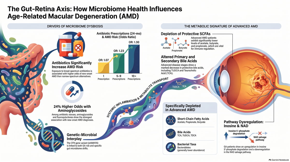
External perturbations such as broad-spectrum antibiotic therapy are significant drivers of long-term gut dysbiosis. Epidemiological data indicates that exposure to specific classes significantly increases the odds of a new-onset GA/AMD diagnosis:
- Aminoglycosides: OR 1.24 (95% CI, 1.22–1.26).
- Fluoroquinolones: OR 1.13 (95% CI, 1.12–1.14).
This association is frequency- and duration-dependent, suggesting that sustained perturbations exacerbate the "gut-retina axis." Fecal metabolomics of advanced AMD cases further elucidates this systemic deficit, showing a depletion of protective metabolites:
- Short-Chain Fatty Acids (SCFAs): Significant decreases are observed in acetate (P=0.002), butyrate (P=0.04), and propionate (P=0.01). SCFAs maintain intestinal barrier integrity; their loss promotes a "leaky gut."
- Bile Acids (BAs): Key primary and secondary BAs are reduced, including taurocholic acid (P=0.02) and tauroursodeoxycholic acid (TUDCA) (P=0.04).
These deficits allow microbial products to enter systemic circulation, triggering the chronic inflammation and complement activation that characterize the GA retina.

7. Clinical Conclusions and Future Investigative Directions
The evidence confirms that Geographic Atrophy is a systemic condition influenced by the Gut-Eye Axis. The pathophysiology involves a stable but functionally altered gut microbiome, a reduction in protective SCFAs and BAs, and a high-risk genetic background centered on the CFH rs1061170 SNP.
Future Research Recommendations:
- Targeting IMPDH: Evaluate the clinical efficacy of IMPDH inhibitors to stabilize retinal nucleotide demand and prevent RPE loss.
- Longitudinal Dysbiosis Studies: Launch prospective studies to track if antibiotic-induced dysbiosis acts as a primary trigger for GA onset in genetically predisposed (C-allele) individuals.
- Microbial Therapeutics: Explore the use of targeted probiotics or metabolic supplementation (NAD precursors or TUDCA) to attenuate retinal degradation.
In summary, the gut microbiome presents a promising diagnostic biomarker for GA progression. However, its greatest potential lies in being a modifiable factor for novel interventions aimed at reducing the systemic inflammatory burden on the retina.


Frequently Asked Questions (FAQs):
How do gut microbiome changes contribute to geographic atrophy pathophysiology?
Gut microbiome changes contribute to geographic atrophy (GA) pathophysiology through a complex multi-step mechanism along the gut-eye (or gut-retina) axis. Although the overall core composition of the gut microbiome remains relatively stable at the phylum level, subtle taxonomic shifts, metabolic pathway alterations, and the depletion of protective gut-derived metabolites drive systemic inflammation, oxidative stress, and complement system activation in retinal tissues.
1. Specific Taxonomic Shifts & Pathobiont Expansion
- Depletion of Protective Species: GA patients exhibit a significant loss of beneficial bacterial species such as Anaerotruncus colihominis and Dorea formicigenerans. The depletion of Anaerotruncus is associated with metabolic dysregulation and altered inflammatory responses, which can exacerbate GA progression by elevating oxidative stress and activating the complement system.
- Pro-inflammatory Shifts: There is a significant upregulation of the genus Firmicutes GGB9758 and increased species abundance of GGB9758 SGB15368 and Gemmiger SGB15299 in GA patients.
- Expansion of Desulfovibrionales & Barrier Dysfunction: Advanced AMD/GA is marked by an increased abundance of the order Desulfovibrionales. These sulfate-reducing bacteria produce toxic hydrogen sulfide (H₂S), which degrades the protective intestinal mucosal layer. This compromised gut epithelial barrier allows endotoxins (such as lipopolysaccharides) to leak into the bloodstream, triggering pro-inflammatory Th1 and Th17 adaptive immune responses that propagate to the eye.
- Clostridia Taxa Accumulation: Advanced disease also features higher abundances of specific families (Peptococcaceae, Gracilibacteraceae) and genera (Butyrivibrio, Lachnotalea, Paludicola, Terrisporobacter) within the Clostridia class, creating a cluster of heightened immune activity linked to retinal damage.
2. Alterations in Functional Metabolic Pathways
Metagenomic functional analysis shows that gut dysbiosis fundamentally alters key biochemical pathways in GA patients:
- Upregulation of Inosine 5′-Monophosphate Degradation: The pathway for inosine 5′-phosphate degradation is significantly upregulated in GA. This pathway involves the enzyme inosine-5′-monophosphate dehydrogenase (IMPDH), which is directly linked to retinal degeneration. Its upregulation disrupts purine nucleotide biosynthesis, depriving the high-demand neuroretina of essential purine nucleotides.
- Downregulation of NAD Salvage Pathways: Both the PNC V and PNC VI NAD salvage cycles are downregulated in GA. Reduced nicotinamide adenine dinucleotide (NAD) levels impair cellular energy metabolism and decrease sirtuin activity (which regulates stress responses and longevity), ultimately elevating oxidative stress in retinal cells.
- Downregulation of Ketogenesis & TCA Pathways: Pathways governing ketogenesis and the reductive TCA cycle II are downregulated. These shifts disrupt mitochondrial energy production and correlate with heightened reactive oxygen species (ROS) production in retinal pigment epithelium (RPE) cells.
3. Depletion of Protective & Neuroprotective Metabolites
- Short-Chain Fatty Acids (SCFAs): Fecal levels of essential SCFAs—including acetate, butyrate, and propionate—are markedly depleted in advanced disease. SCFAs normally maintain gut barrier tightness, promote microglial metabolic fitness, alter autoreactive T-cell trafficking, and exert direct anti-inflammatory and anti-angiogenic effects on retinal tissues.
- Bile Acids (BAs): Primary and secondary conjugated bile acids, such as taurocholic acid (TCA), taurochenodeoxycholic acid (TCDCA), and tauroursodeoxycholic acid (TUDCA), are significantly decreased in GA/advanced AMD. These molecules normally promote RPE cell integrity, inhibit NF-κB-mediated inflammation, reduce oxidative damage, and protect photoreceptor function.
4. Interactions with Host Genetics & Environmental Triggers
- Genetic Risk Modulation (rs1061170): Carrying the high-risk C allele of SNP rs1061170 in the complement factor H (CFH) gene is strongly associated with GA (OR = 2.94). GA patients with this genetic risk show a distinct upregulation of the gut bacterial genus Anaerotignum, demonstrating how host complement genetics modulate gut microbiota composition to drive systemic inflammatory signaling.
- Antibiotic-Induced Dysbiosis: Cumulative exposure to broad-spectrum antibiotics, tetracyclines (e.g., doxycycline), and quinolones is associated with increased odds of developing GA in a dose-dependent manner. Antibiotics disrupt microbial diversity and damage the intestinal mucosal barrier, initiating chronic inflammation that disseminates along the gut-retina axis to accelerate RPE and photoreceptor degeneration.
How do SCFA and bile acid supplements help?
Supplements of Short-Chain Fatty Acids (SCFAs) (such as acetate, butyrate, and propionate) and bile acids (such as TUDCA, TCA, and TCDCA) help counter geographic atrophy (GA) and advanced age-related macular degeneration (AMD) by targeting systemic inflammation, intestinal barrier breakdown, microglial dysfunction, and retinal cell stress along the gut-retina axis.
1. Protective Mechanisms of Short-Chain Fatty Acid (SCFA) Supplementation
Fecal levels of key SCFAs—including acetate, butyrate, and propionate—are depleted in patients with advanced retinal degeneration. Supplementing SCFAs provides protection through several distinct pathways:
- Restoring Gut Barrier Integrity & Immune Homeostasis: SCFAs reinforce the intestinal epithelial lining and decrease gut permeability, preventing microbial endotoxins (like LPS) from leaking into circulation. Systemically, oral SCFA administration alters the trafficking of autoreactive effector T cells and induces protective regulatory T cells (Tregs), thereby suppressing chronic ocular inflammation.
- Sustaining Microglial Fitness: Retinal and CNS microglia rely on acetate for metabolic fitness, phagocytic clearance, and proper maturation. Replenishing acetate levels helps prevent the microglial dysfunction characteristic of neurodegenerative states.
- Inhibiting Pathological Angiogenesis: SCFAs like sodium butyrate exert direct anti-angiogenic and anti-inflammatory actions in ocular tissue by downregulating VEGFA and VEGFR2 signaling pathways, thereby limiting vascular and retinal pigment epithelium (RPE) damage.
2. Protective Mechanisms of Bile Acid Supplementation
Conjugated primary and secondary bile acids act as signaling molecules across the gut-retina axis, but their concentrations drop significantly as disease severity progresses. Supplementation restores key biochemical defenses:
- Promoting RPE Cell Integrity: Taurocholic acid (TCA) enhances the structural integrity of RPE cells and decreases VEGF-induced choroidal endothelial cell migration.
- Suppressing Neuroinflammation via TGR5 & NF-κB: Taurochenodeoxycholic acid (TCDCA) suppresses inflammatory gene expression—including TNF-α, IL-6, and COX-2—by activating TGR5 receptors and inhibiting pro-inflammatory NF-κB signaling.
- Neuroprotection & ER Stress Reduction (TUDCA): Tauroursodeoxycholic acid (TUDCA) is a neuroprotective bile acid that attenuates endoplasmic reticulum (ER) stress, rescues cone photoreceptors from cell death, protects microvascular endothelial cells, and preserves overall retinal structural function.
-
Regulating Lipid & Energy Homeostasis: Conjugated bile acids (such as tauro-α/β-muricholic acid) serve as natural farnesoid X receptor (FXR) antagonists, regulating systemic lipid metabolism and energy pathways involved in chronic inflammation.
Could you explain the role of TUDCA in neuroprotection?
Tauroursodeoxycholic acid (TUDCA) is a conjugated secondary bile acid that acts as a potent neuroprotective and retinoprotective signaling molecule. In patients with advanced age-related macular degeneration (AMD), fecal concentrations of TUDCA are significantly depleted compared to both healthy controls and patients with intermediate AMD.
The core mechanisms through which TUDCA exerts its neuroprotective and eye-protective effects include:
- Cone Photoreceptor Rescue & Functional Protection: TUDCA directly rescues cone photoreceptors in degenerative retinas. In animal models of retinal degeneration (such as rd1 mice), TUDCA preserves both retinal structural integrity and visual function.
- Attenuation of Endoplasmic Reticulum (ER) Stress: TUDCA attenuates age-related and diet-induced endoplasmic reticulum (ER) stress, a fundamental cellular stress pathway implicated in neurodegenerative conditions and retinal cell death.
- Reduction of Amyloid Accumulation: TUDCA targets age-dependent neurodegenerative processes by reducing the accumulation of amyloid deposits.
- Endothelial Regulation & Retinopathy Protection: TUDCA reduces human endothelial cell proliferation and protects against retinal microvascular endothelial dysfunction, diabetic retinopathy, and broader retinal degeneration.
Together, these findings highlight TUDCA as a vital protective metabolite along the gut-retina axis; its loss in advanced AMD removes a critical defense against ER stress, amyloid toxicity, and photoreceptor loss.
How do Mediterranean diets boost SCFAs?
- Fermentable Substrates: The Mediterranean diet emphasizes plant-based whole foods, including legumes, vegetables, fruits, whole grains, nuts, and seeds.
- Colonic Fermentation: These foods are rich in complex dietary fibers and resistant starches (such as inulin, pectins, and cellulose) that resist digestion in the upper gastrointestinal tract. Upon reaching the colon, commensal anaerobic bacteria—such as Faecalibacterium prausnitzii, Bacteroides species, and key taxa within the Lachnospiraceae and Ruminococcaceae families—use these fibers as fuel, fermenting them directly into SCFAs.
- Polyphenol-Driven Microbial Selection: Extra virgin olive oil, nuts, seeds, and colorful plant foods are abundant in polyphenols. These bioactive compounds act as prebiotics that selectively promote the proliferation of SCFA-producing commensals while suppressing pro-inflammatory bacteria.
- Gut Barrier Protection: Polyunsaturated and monounsaturated fatty acids support gut mucosal integrity, providing a stable anaerobic microenvironment necessary for fermentative SCFA synthesis.
- Reduction of Western Diet Elements: Unlike high-fat, high-glycemic Western diets—which nourish sulfate-reducing bacteria (like Desulfovibrionales) and deplete anaerobic SCFA producers—the Mediterranean diet avoids refined sugars and excessive saturated fats.
- Sustained Microbial Fitness: Minimizing refined sugars prevents chronic gut dysbiosis, ensuring that fiber-fermenting bacterial populations remain dominant and capable of maintaining optimal luminal SCFA concentrations.
Are there targeted prebiotic supplements for GA?
There are currently no targeted or FDA-approved prebiotic supplements specifically formulated or clinically validated for Geographic Atrophy (GA).
While prebiotic supplementation specifically designed for GA does not yet exist in clinical practice, current research provides a strong rationale for why gut-directed therapies are being investigated:
1. Current Clinical Evidence for Dietary Interventions
- Proven Formulations: To date, the established nutritional strategy for reducing the risk of progression in appropriate patients with AMD involves formulations containing antioxidants, lutein, zeaxanthin, and other nutrients, such as the AREDS2 regimen.
- Lack of GA-Specific Prebiotic Trials: Clinical studies evaluating prebiotics—non-digestible fibers that feed beneficial bacteria—specifically for halting or preventing GA progression have not yet been conducted.
2. The Biological Basis for Prebiotic Research in GA
Although targeted products are not currently available, researchers are studying the gut-retina axis because of distinct microbial and metabolic differences observed in GA:
- Restoring Depleted Metabolites: Patients with advanced AMD and GA exhibit depletions in gut-derived short-chain fatty acids (SCFAs)—such as acetate, butyrate, and propionate—as well as potentially protective bile acidssuch as TUDCA and TCA. In theory, prebiotic substrates such as inulin, pectin, or resistant starches could nourish fermenting bacteria and influence these metabolite pools.
- Countering Taxonomic Dysbiosis: GA has been associated with a loss of potentially protective species, such as Anaerotruncus colihominis and Dorea formicigenerans, alongside increased abundance of potentially pro-inflammatory taxa, including Desulfovibrionales and specific Clostridia. Prebiotics are being investigated more broadly for their potential to selectively promote beneficial commensal bacteria and influence microbial balance.
Important distinction: These findings provide a research rationale, not evidence that taking a prebiotic supplement prevents, treats, or slows GA. Clinical trials would be needed to establish whether modifying the gut microbiome produces meaningful benefits for patients with GA.
Anaerotruncus colihominis and Dorea formicigenerans)?
- Significant Depletion in GA: Whole-metagenome shotgun sequencing shows that Anaerotruncus colihominis is markedly less abundant in the gut microbiome of GA patients compared to age- and sex-matched healthy controls.
- Metabolic & Gastrointestinal Function: The genus Anaerotruncus is involved in normal intestinal metabolism and maintaining gastrointestinal homeostasis.
-
Impact on GA Pathophysiology:
- Systemic Dysregulation: Its depletion is linked to altered metabolic pathways and impaired anti-inflammatory control.
- Oxidative Stress & Complement Activation: Loss of Anaerotruncus contributes to a pro-inflammatory systemic environment, elevating oxidative stress levels and triggering complement system hyperactivation—two major drivers of retinal pigment epithelium (RPE) damage and photoreceptor loss in GA.
- Significant Downregulation in GA: Dorea formicigenerans is significantly downregulated in patients with GA relative to healthy individuals.
- Commensal Role: Dorea species are key fermenting organisms in the human gut that contribute to metabolic regulation, dietary carbohydrate breakdown, and the maintenance of a balanced gut microenvironment.
-
Impact on GA Pathophysiology:
- Metabolic Imbalance: Because Dorea formicigenerans is closely tied to metabolic and immune signaling, its depletion impairs normal microbial fermentative activity.
- Gut-Retina Signaling: Decreased abundance of this species correlates with broader functional pathway dysregulation—such as shifts in purine nucleotide degradation and reduced energy pathway activity—which weakens endogenous defenses against chronic ocular inflammation.
- High-Fiber & Plant-Rich Diets: Diets rich in complex carbohydrates, resistant starches, and non-digestible fibers (found in legumes, whole grains, vegetables, and seeds) serve as primary fermentative fuel. Commensal species use these fibers to produce protective SCFAs like acetate, butyrate, and propionate.
- Polyphenols & Monounsaturated Fats: Foods rich in polyphenols (such as extra virgin olive oil, nuts, and berries) act as natural prebiotics. They selectively foster a favorable gut environment for beneficial species while suppressing pro-inflammatory, sulfate-reducing bacteria.
- Reducing High-Glycemic & High-Saturated-Fat Foods: Minimizing Western-style diet components prevents the overgrowth of pathobionts that disrupt the mucosal barrier and outcompete beneficial fermenting taxa.
- Avoiding Unnecessary Broad-Spectrum Exposure: Broad-spectrum antibiotics, fluoroquinolones, and tetracyclines significantly disrupt gut microbial diversity and cause long-term depletions of anaerobic commensal bacteria.
- Targeted Prescribing: Minimizing cumulative antibiotic duration and choosing narrow-spectrum agents when clinically appropriate helps preserve core commensal taxa and prevents prolonged dysbiosis.
- Prebiotic Supplementation: Non-digestible oligosaccharides (such as inulin, fructooligosaccharides, and galactooligosaccharides) can be used to selectively stimulate the growth and metabolic activity of depleted bacterial families.
- Next-Generation Biotherapeutics: While traditional commercial probiotics focus primarily on Lactobacillus and Bifidobacterium strains, research is actively developing next-generation live biotherapeutic products (LBPs) that incorporate specialized anaerobic commensals to directly reseed depleted gut ecosystems.
- SCFA Supplementation: Oral or targeted formulations of butyrate, acetate, and propionate can directly reinforce the intestinal epithelial barrier, reduce systemic inflammation, and support retinal cell health.
- Bile Acid Restoration: Supplementing neuroprotective bile acids like TUDCA helps counteract the functional deficit caused by missing microbial taxa, protecting photoreceptors and reducing endoplasmic reticulum stress along the gut-retina axis.
What is the 'gut-retina axis'?
A hypothesized link where gut microbiome dysbiosis and resulting metabolic changes contribute to the pathogenesis of retinal diseases.
Which antibiotic class is associated with a 24% increase in the odds of a new-onset diagnosis of age-related macular degeneration (AMD)? Aminoglycosides.
How does the risk of new-onset AMD change with the cumulative number of antibiotic prescriptions? The risk increases in a frequency-dependent manner, with ten or more prescriptions showing the highest odds (OR=1.30).
Exposure to which antibiotic class is associated with an 18% increase in the odds of new-onset Geographic Atrophy (GA)? Tetracyclines.
How does broad-spectrum antibiotic exposure compare to narrow-spectrum exposure regarding the odds of new-onset AMD? Broad-spectrum antibiotics are associated with nearly three times greater odds (OR=1.15 vs 1.05).
According to administrative claims data, a cumulative antibiotic supply of ≥57 days increases the odds of new-onset Geographic Atrophy (GA) by? 38%
How do fluoroquinolones like ciprofloxacin potentially damage the retina at the cellular level? They decrease the viability of retinal pigment epithelium (RPE) cells and upregulate pro-apoptotic genes.
In the gut microbiome of Geographic Atrophy (GA) patients, what is the status of the NAD salvage pathway? It is significantly downregulated
The inosine 5-phosphate degradation metabolic pathway is significantly upregulated in the gut microbiome of patients with Geographic Atrophy.
Which gut bacterial genus shows an upregulation in Geographic Atrophy (GA) patients who are homozygous for the SNP rs1061170? Anaerotignum.
Why are broad-spectrum antibiotics hypothesized to carry a higher risk for AMD development than narrow-spectrum antibiotics? They cause more severe perturbations and larger deviations from the gut microbiome's homeostatic baseline.
Advanced AMD patients show a significant decrease in the abundance of which three short-chain fatty acids (SCFAs)? Acetate, butyrate, and propionate.
Which metabolic cycle, associated with the production of reactive oxygen species (ROS), is downregulated in the gut microbiome of GA patients? The reductive TCA cycle.
How does the association between quinolone exposure and 'wet' AMD compare to its association with 'dry' AMD?
The association is significantly stronger for wet AMD, exhibiting higher odds ratios.
Exposure to which two commonly prescribed oral antibiotics is associated with a 19% increase in the odds of new-onset Geographic Atrophy? Cephalexin and doxycycline.
t what time interval before diagnosis is fluoroquinolone exposure associated with the highest odds of a new-onset AMD diagnosis? Zero to six months.
The process where dysbiosis enables the translocation of microbial endotoxins into systemic circulation is thought to trigger the inflammation observed in AMD.
How does the impact of standard-dose doxycycline (100−200 mg) on GA risk contrast with its proposed sub-antimicrobial therapeutic use? Standard-dose doxycycline is associated with increased GA risk, whereas low-dose doxycycline (40 mg) is being studied to slow disease progression.
Advanced AMD patients exhibit a significant decrease in which specific bile acid that inhibits AMD features in vitro? Taurocholic acid.
What is the primary characteristic of the ocular surface microbiome (OSM) in Geographic Atrophy (GA) patients compared to controls? The OSM shows no significant taxonomic shifts or differences between the two groups.
Which bacterial genus linked to immune activity and rheumatoid arthritis is found in greater abundance in advanced AMD compared to intermediate AMD Terrisporobacter.
The downregulation of which metabolic pathway in the GA gut microbiome indicates changes in energy metabolism linked to oxidative stress? Ketogenesis.
Which specific antibiotic class is associated with a 16% increase in the odds of new-onset Geographic Atrophy? Quinolones.
How do short-chain fatty acids (SCFAs) like butyrate generally affect gut health relevant to the gut-retina axis? They act as anti-inflammatory mediators and help reduce intestinal permeability.
Which enzyme involved in nucleotide biosynthesis is suggested as a potential therapeutic target due to its upregulation in GA pathophysiology? Inosine-5'-monophosphate dehydrogenase (IMPDH).
In the ocular surface microbiome of GA patients, disruptions in which enzyme pathway may lead to tear film instability? Phospholipases.
What is the median day supply of antibiotics for cases of new-onset Geographic Atrophy compared to controls? Twelve days for cases versus ten days for controls.
Besides smoking and diet, antibiotic use is now being investigated as a novel modifiable risk factor for Geographic Atrophy.
Does Age Influence Antibiotic-Associated Macular Degeneration Risk?
1. Age as a Factor in Antibiotic-Associated AMD and GA Risk
In the large national studies evaluating antibiotic exposure and macular degeneration, patient age was controlled for by matching cases and controls 1-to-1 by age.
- Study Population Ages: The general AMD study focused on adults aged 55 and older (mean age 74.9 years), whereas the Geographic Atrophy (GA) study focused on adults aged 60 and older (mean age 81.0 years).
- Independent Risk: Because cases and controls were matched by age, the increased odds of developing AMD or GA from cumulative antibiotic use were observed independently of patient age.
- Distribution Across Age Groups: For GA, new-onset cases occurred across all evaluated age groups: 15.2% in patients aged 60–69, 25.8% in those aged 70–79, 37.2% in those aged 80–89, and 21.9% in those aged 90 and older.
2. Effect of Taking Antibiotics at a Younger Age
- Unmeasured in Adult Claims Studies: The AMD and GA claims studies evaluated antibiotic prescriptions within a 24-month lookback window before diagnosis, so early-life or lifetime antibiotic exposures were not captured.
- Hypothesized Cumulative Dysbiosis: The researchers highlighted that measuring early-life or lifetime antibiotic use is an important area for future study, as early perturbations to the microbiome may trigger sustained gut dysbiosis and chronic low-grade inflammation that persist over decades.
- Contrast with Younger Cohorts: A smaller study in a younger population (mean age 49.8 years) with only a 1-month follow-up found no short-term retinal changes from fluoroquinolones. This suggests that underlying age-related tissue vulnerability, combined with a sufficient time window for chronic inflammatory changes to develop, is necessary for antibiotic-associated AMD or GA to manifest.
- Early-Life Impact in Ocular Disease: Although younger-age exposure was not tracked for adult AMD, early-life exposure in infants has been studied in other retinal conditions. Specifically, exposure to broad-spectrum antibiotics within the first two months of life in preterm infants significantly increased the odds of developing severe Retinopathy of Prematurity (ROP) through early gut dysbiosis.

Leave a comment
This site is protected by hCaptcha and the hCaptcha Privacy Policy and Terms of Service apply.