
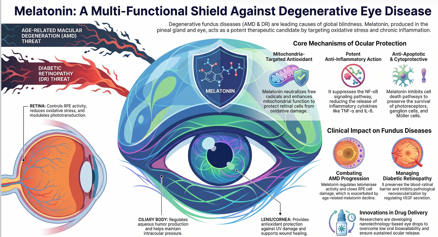
Abstract: Age-related macular degeneration (AMD) remains a leading cause of irreversible visual impairment worldwide. Recent translational and epidemiological research has reshaped our understanding of AMD pathobiology, shifting focus from passive debris accumulation to an active, self-amplifying cycle of mitochondrial degradation, lipid-driven regulated cell death, and immune overactivation. Central to this network is the progressive loss of retinal pigment epithelium (RPE) cells and macular cones. Excitingly, melatonin—synthesized both in the pineal gland and locally within the neuroretina—emerges as a potent chorioretinal shield. Beyond its classical role as a chronobiotic hormone, melatonin operates as an intra-mitochondrial radical scavenger and an upstream epigenetic and transcriptional regulator. This review synthesizes the molecular mechanisms by which melatonin preserves retinal bioenergetics, regulates autophagic flux, and suppresses ferroptosis and inflammasome assembly. Furthermore, we evaluate real-world human cohort data and next-generation nanotechnological ophthalmic delivery architectures that bridge the gap between bench discovery and non-invasive clinical prevention.

1. Introduction: The Bioenergetic Crisis in Retinal Aging
The human fovea centralis possesses one of the highest metabolic rates of any tissue in the body, driven by the ceaseless ATP demands of phototransduction and dark adaptation. To sustain this exceptional metabolic load, the neuroretina and the underlying retinal pigment epithelium (RPE) monolayer are packed with mitochondria. This extreme bioenergetics environment operates under a permanent state of oxidative threat. The RPE is continuously exposed to high oxygen tension from the choriocapillaris, focused visible light, and easily oxidizable polyunsaturated fatty acids (PUFAs) derived from the daily phagocytosis of photoreceptor outer segments.
With advanced age and environmental insults (e.g., cigarette smoke, phototoxic blue-light exposure, and ambient volatile organic compounds), this delicate redox balance collapses. The aging chorioretinal interface experiences a progressive decline in endogenous free-radical scavenging enzymes, culminating in an accumulation of reactive oxygen and nitrogen species. This oxidative stress damages mitochondrial components, leading to a self-perpetuating bioenergetic crisis. Over the past decade, a major paradigm shift has repositioned this mitochondrial decay as the primary driver of dry AMD and geographic atrophy, preceding overt clinical tissue loss.
Simultaneously, the neurohormone melatonin has emerged as an indispensable local homeostatic factor in the retina. While long celebrated as the systemic master signal of darkness, melatonin is synthesized locally within retinal photoreceptors and exhibits unique intra-mitochondrial properties. Crucially, clinical profiling reveals that patients suffering from AMD exhibit significantly lower systemic and local melatonin levels compared to age-matched controls, confirming that a breakdown in melatoninergic signaling is tightly interwoven with macular degeneration. This review explores the multi-tiered protective mechanisms of melatonin in the aging retina, providing a mechanistic and clinical rationale for its development as a targeted ophthalmic therapy.

2. Preclinical & Molecular Pathobiology of Dry AMD
The progressive degeneration of RPE cells in geographic atrophy follows a sophisticated, multi-pathway destructive cascade initiated by genetic vulnerability and catalyzed by mitochondrial failure.
The Disease Progression Cascade:
- DICER1 Deficiency leads to Alu RNA Accumulation.
- This forces mitochondrial pore opening and mitochondrial DNA leakage into the cell.
- Cytoplasmic DNA activates cGAS-STING and the NLRP3 Inflammasome.
- Inflammasomes trigger IL-18 Production.
- Autocrine signaling forces Caspase-8 Apoptosis (Cell Death).

2.1. The DICER1/Alu RNA Toxic Loop and Inflammasome Priming
Geographic atrophy is fundamentally characterized by a prominent deficiency of the microRNA-processing enzyme DICER1 within the RPE monolayer. Beyond its role in microRNA biogenesis, DICER1 functions to cleave the non-coding Alu retrotransposon element. Its deficiency causes an overwhelming accumulation of Alu RNA transcripts.
This Alu RNA excess acts as a potent intracellular toxin that directly coordinates mitochondrial collapse:
- It triggers the permanent opening of mitochondrial permeability transition pores.
- It induces a profound loss of mitochondrial membrane potential.
- It causes the extrusion of mitochondrial DNA into the cytoplasm.
Once in the cytoplasm, the displaced mitochondrial DNA interacts with cyclic GMP-AMP synthase (cGAS), stimulating the downstream cGAS-STING pathway to drive non-canonical activation of the NLRP3 inflammasome. The fully assembled NLRP3 inflammasome activates caspase-1, leading to the maturation and secretion of interleukin-18 (IL-18). Operating in a destructive autocrine loop, IL-18 triggers internal dependent signaling that culminates in caspase-8-mediated RPE cell apoptosis. This inflammatory, cell-lethal cascade is amplified by extracellular drusen components—including amyloid-beta, complement fragments, and the phototoxic bisretinoid A2E—which concurrently prime the NLRP3 complex.

2.2. Superoxide-Driven Ferroptosis via the Haber-Weiss Reaction
While apoptosis handles a fraction of RPE death, recent evidence establishes iron-dependent, lipid-peroxidation-driven ferroptosis as the predominant executioner of light-induced outer retinal damage. Mitochondria produce substantial amounts of superoxide anions, primarily via electron leakage at respiratory complexes I and III. Under healthy conditions, manganese superoxide dismutase (MnSOD/SOD2) in the mitochondrial matrix converts these toxic anions into hydrogen peroxide for downstream enzymatic neutralization.

In the context of AMD, an age-dependent or light-induced drop in MnSOD activity allows superoxide to accumulate. This excess superoxide initiates an iron-catalyzed loop through the Haber-Weiss reaction, where superoxide anions interact with ferric iron to generate highly destructive hydroxyl radicals directly within the lipid-dense mitochondrial membranes. These radicals attack nearby polyunsaturated fatty acids, inducing rampant lipid peroxidation marked by the accumulation of malondialdehyde (MDA) and 4-hydroxynonenal (4-HNE).
Compounding this crisis, the resulting mitochondrial reactive oxygen species overproduction up-regulates the molecular chaperone Heat Shock Protein 70 (Hsp70). Hsp70 directly binds to glutathione peroxidase 4 (GPX4)—the master surveillance enzyme responsible for neutralizing lipid peroxides—and targets it for ubiquitin-proteasome degradation. Deprived of GPX4, the RPE cell undergoes irreversible ferroptotic membrane rupture, triggering localized geographic atrophy.

3. Retinal Melatonin Chronobiology and Receptor Dynamics
The mammalian retina does not merely absorb light for image formation; it acts as a highly integrated, light-sensitive clock that relies on melatonin as its core nocturnal mediator.
3.1. Synthesis, Local Rhythms, and the Dopamine Axis
Retinal melatonin is synthesized locally within photoreceptor cells via an autonomous molecular clock that operates independently of the master brain pathways. Retinal melatonin production exhibits a high-amplitude daily rhythm, peaking sharply at night and falling to near-zero levels during daylight hours. This temporal gating is achieved via a dual mechanism:
- Transcriptional Control: The autonomous photoreceptor clock drives the rhythmic transcription of the rate-limiting enzyme arylalkylamine N-acetyltransferase (AANAT) and adenylate cyclase 1 (Adcy1).
- Post-Translational Control: At night, elevated cellular messengers trigger protein kinase A (PKA)-mediated phosphorylation of AANAT, stabilizing the enzyme against prosomal degradation.
Conversely, daytime light exposure activates dopamine synthesis and release from dopaminergic amacrine cells. Dopamine binds to specific receptors on photoreceptors, suppressing internal signaling and causing rapid dephosphorylation and proteasomal clearing of AANAT, effectively quenching melatonin production. Retinal melatonin thus operates in permanent, reciprocal antagonism to daytime dopamine.

3.2. Receptor Signaling and Outer Retinal Plasticity
Melatonin executes its localized, intraocular functions by binding to two high-affinity, seven-transmembrane G-protein-coupled receptors, MT1 and MT2, which are heavily expressed across the neuroretina and RPE. In the outer retina, MT1 and MT2 physically assemble into functional receptor heteromers on photoreceptor membranes.
This receptor network directly shapes retina circuitry and dark adaptation through the modulation of rod/cone gap junctions. During daylight, high dopamine levels keep the gap junction protein Connexin36 (Cx36) phosphorylated, decoupling rods from cones. At night, the surge of melatonin suppresses dopamine, lowering PKA activity and increasing the open probability of Cx36 channels. This increases rod/cone electrical junctional conductance from a daytime baseline of 0–50 pS to a nighttime maximum of 1200 pS, injecting dim-light rod signals directly into cone visual pathways to maximize visual light sensitivity.
4. Melatonin as an Intra-Mitochondrial Shield
Beyond its classical membrane receptor-mediated functions, melatonin exhibits exceptional, direct cytoprotective capabilities that stem from its unique evolutionary origin as an intra-mitochondrial antioxidant.
Summary of Melatonin's Role in Protecting Mitochondria
- Mitochondrial Uncoupling: High membrane potential drives electron leakage and excessive free radical production. Melatonin upregulates and activates Uncoupling Proteins (UCP2), safely dissipating the internal gradient to limit baseline free radical generation.
- Epigenetic Regulation: Age-related drops in downstream synthesis parameters damage the outer blood-retinal barrier. Melatonin upregulates specific demethylase pathways, which recruits protective regulatory T cells and switches microglia from a pro-inflammatory M1 state to a healing M2 state.
- Bioenergetic Rescue: Specific enzymatic inhibition typically causes a loss of active mitochondrial mass. Melatonin can donate and accept electrons directly in the electron transport chain, stabilizing tracking channels and protective proteins to restore ATP synthesis and preserve mitochondrial mass.
- Autophagic Flux: Autophagy arrest leads to an accumulation of toxic cellular debris. Melatonin suppresses negative-feedback phosphorylation pathways, which helps convert internal structural components, increases Beclin-1, and clears toxic protein aggregates.
- Ferroptosis Surveillance: Excess reactive oxygen species trigger the proteasomal decay of surveillance enzymes. Melatonin eliminates these free radicals via the main cellular metabolic pathways, preventing structural upregulation of damaging chaperones and saving protective pathways from degradation.
Mitochondria are the primary site of melatonin synthesis and accumulation within cells. Melatonin is actively concentrated inside the mitochondrial matrix against its gradient via special peptide transporters localized on the inner membrane. Once inside, melatonin serves as a highly efficient, self-regenerating free-radical scavenger. It neutralizes hydroxyl, peroxyl, and singlet oxygen radicals via a continuous detoxification cascade where its metabolic by-products retain equal or superior antioxidant capacity.

Concurrently, melatonin acts via an integrated transcriptional network. By stimulating upstream indicators, melatonin upregulates protective master sirtuin (SIRT3) expression in the matrix. SIRT3 directly activates MnSOD, accelerating the dismutation of superoxide anions and blocking the Haber-Weiss reaction before it can synthesize lethal hydroxyl radicals. Furthermore, melatonin directly protects mitochondrial DNA from incorporating point mutations, preserves membrane lipid structures, and maintains inner membrane stability. This multi-layered mechanical support prevents mitochondrial pore opening, blocking the exit of death effectors into the cytoplasm, thereby intercepting RPE cell death.

5. Clinical & Real-World Evidence of Melatonin Efficacy
突然oving beyond preclinical configurations, recent large-scale clinical and real-world database mining provides robust confirmation of melatonin's efficacy in protecting the human retina.

5.1. The TriNetX Big Data Cohort Analysis
The most mathematically sound evidence validating melatonin's real-world protective capacity was delivered in a massive retrospective cohort study utilizing the TriNetX healthcare network. This study extracted deidentified electronic health records from over 95 million patients across more than 60 US healthcare organizations. To control for health status, lifestyle, and healthy-user biases, cohorts were subjected to strict propensity score matching (PSM), balancing demographics, smoking status, systemic comorbidities, and non-melatonin hypnotic medication usage.

The comparative analysis targeted two primary outcomes:
- The development of new, de novo AMD diagnoses in patients with no prior history of the disease.
- The progression of preexisting nonexudative (dry) AMD to exudative (wet) AMD.
The results demonstrated a highly significant, negative association between regular melatonin use and AMD outcomes across all age groups:
- In the AMD-naive cohort (aged 50 years or older), melatonin usage was associated with a 58% reduction in the risk of developing new AMD (Risk Ratio: 0.42; 95% Confidence Interval: 0.28–0.62). This preventative effect intensified in older brackets, achieving a 65% risk reduction in patients aged 70 years or older (Risk Ratio: 0.35; 95% Confidence Interval: 0.23–0.53).
- In patients with preexisting nonexudative AMD, melatonin use lowered the rate of conversion to the aggressive exudative (wet) phenotype by 56% (Risk Ratio: 0.44; 95% Confidence Interval: 0.34–0.56). This containment remained highly significant even after filtering out patients receiving active anti-growth factor intraocular injection therapy.
- Crucially, when patients were monitored for long-term outcomes occurring at least 2 years after the initial event, melatonin's preventative efficacy increased further, achieving up to a 78% reduction in new AMD risk (Risk Ratio: 0.22).

5.2. Human Electrophysiological Outcomes
This real-world epidemiological protection correlates with objective functional improvements in human retinal signaling. In clinical trials exploring early-to-advanced degenerative conditions, oral melatonin supplementation (2–3 mg nightly) stabilized visual acuity and halted progressive macular changes in a majority of treated subjects.
To quantify this protection electrophysiologically, researchers have utilized pattern electroretinography to monitor inner retinal health. Glaucoma and macular degeneration patients treated with regular melatonin demonstrated a significant morning increase in the amplitudes of the pattern electroretinogram N95 wave—a highly sensitive wave that originates explicitly from the retinal ganglion cells and optic nerve. This functional optimization reflects enhanced cellular resilience and retinal transmission efficiency within the macula, verifying that melatonin actively rejuvenates failing neural pathways in the human eye.
 6. Breakthroughs in Ophthalmic Nano-Formulation and Delivery
Formulating melatonin as eye drops presents two primary challenges: poor water solubility and rapid precorneal elimination driven by the protective barriers of the ocular surface. Standard eye drops exhibit an ocular bioavailability of less than 5%, necessitating the engineering of advanced nanotechnological delivery systems to facilitate stable, transcorneal diffusion.
The Delivery Strategy: Topical Drops -> Encapsulation in Nanomicelle / Cationic Cores -> Prolonged Precorneal Retention -> Transcorneal / Trans-Scleral Permeation -> Slow Release directly to Retinal Tissue.
6.1. Polymeric Nanomicelles
To transcend solubility limits without using toxic organic solvents, researchers developed an innovative polymeric nanomicellar platform utilizing Soluplus® (a graft copolymer that forms micelles in aqueous solutions). Because of its low critical micellar concentration, the system spontaneously assembles into highly stable, ultra-small nanomicelles (diameter under 200 nm) in an aqueous environment.
This advanced structure encapsulates hydrophobic melatonin inside its core, elevating its aqueous concentration up to 17.2 mM (0.4% w/v). When applied topically, these nanomicelles optimize corneal residence time and facilitate vertical and lateral diffusion pathways through the cornea, aqueous humor, and vitreous body. In hypertensive glaucoma and retinopathy models, this nanomicellar vehicle achieved a 60% reduction in intraocular pressure and significantly enhanced retinal ganglion cell survival, outperforming standard medications like timolol or brimonidine.
6.2. Surface-Functionalized Cationic Nanocarriers
Another breakthrough comprises the fabrication of hybrid polymeric nanocapsules and solid lipid nanoparticles (SLNs) surface-functionalized with cationic lipids, such as didodecyldimethylammonium bromide (DDAB). DDAB coating builds a strongly positive surface charge on the nanocarrier shell. This positive charge maximizes electrostatic interaction with the negatively charged mucin layer of the ocular surface, creating a bioadhesive reservoir that resists nasolacrimal washout.
These functionalized nanosystems demonstrate an encapsulation efficiency exceeding 87% to 96% and follow a controlled, slow-release profile. In rabbit models of retinal degeneration, topical instillation of these cationic nanocapsules safely crossed the cornea and delivered therapeutic concentrations directly to the posterior pole, providing robust structural preservation of the outer retinal layers without inducing ocular surface irritation.

7. Conclusions and Future Directions
The integration of advanced molecular pathobiology, nationwide human epidemiological data, and nanotechnological innovations establishes melatonin as a cornerstone of next-generation multi-target therapies for age-related macular degeneration. AMD is no longer viewed as an unmodifiable consequence of chronological aging, but rather as a manageable bioenergetic failure. Melatonin cuts to the root of this failure by stabilizing mitochondrial respiratory chain complexes, restoring defective autophagic flux, and suppressing the superoxide-driven ferroptotic and inflammasome loops that destroy RPE cells. The real-world clinical protection verified by big data cohort mining—reducing de novo AMD incidence by up to 65%—confirms the high translational readiness of this neuroprotective agent.


To fully maximize melatonin's clinical utility, future research must prioritize three areas:
- Standardization of Biomarker Panels: Longitudinal quantification of oxidative stress footprints (such as urinary melatonin ratios, cell structure intensity, and aqueous humor values) must be standardized to accurately stratify patients.
- Resolution of the Translational Formulation Gap: Ophthalmic clinical practice must transition from systemic oral supplements to targeted, non-invasive topical eye drops. This transition requires industrial scaling and long-term stability testing of mucoadhesive cationic nanocapsules and polymeric nanomicelles capable of delivering consistent, slow-release therapeutic concentrations directly to the macular microenvironment.
- Rigorous Randomized Clinical Trials: Large-scale, multicenter trials utilizing high-resolution structural and functional endpoints are required to define optimal, genotype-specific dosing windows.


By successfully bridging these laboratory breakthroughs with targeted industrial engineering, modern vision science can capitalize on evolution’s premier antioxidant molecule to defend chorioretinal bioenergetics, halt geographic atrophy, and preserve human sight into advanced age.


Frequently Asked Questions (FAQs):
1. What is Age-Related Macular Degeneration (AMD) and how does it affect vision? Age-related macular degeneration (AMD) is a leading cause of irreversible central vision loss among people over the age of 50. It affects the macula, the central part of the retina responsible for sharp, straight-ahead vision. AMD is categorized into two main types: dry AMD, characterized by the accumulation of lipid deposits called drusen and the progressive loss of retinal cells, and wet AMD, which involves the growth of abnormal, leaky blood vessels under the retina.
2. Can melatonin help prevent or treat macular degeneration? Yes, emerging research shows that melatonin supplementation may be highly beneficial for AMD. A large-scale clinical database study involving over 120,000 patients revealed that melatonin use reduced the risk of developing AMD by 58%. Furthermore, for patients who already had dry AMD, melatonin was associated with a 57% decreased risk of the disease progressing to the more severe wet form.
3. How does melatonin protect the eyes from macular degeneration? Melatonin acts as a powerful, multi-targeted protector for the eyes. It is a highly effective antioxidant that scavenges toxic free radicals and protects retinal cells from oxidative stress and lipid peroxidation. Additionally, melatonin protects mitochondrial function within the retina, suppresses chronic inflammation, and inhibits the pathological growth of blood vessels (angiogenesis) that causes wet AMD.
4. Is there a link between sleep disorders, like insomnia, and macular degeneration? Yes, clinical studies indicate a strong association between disrupted circadian rhythms, such as clinically diagnosed insomnia, and an elevated risk of developing AMD. Because melatonin regulates the sleep-wake cycle and acts as a powerful ocular antioxidant, decreased nocturnal melatonin production and poor sleep efficiency can leave the retina vulnerable to oxidative damage and accelerate macular degeneration.
5. Does melatonin work for both "dry" and "wet" macular degeneration? Yes, melatonin targets the underlying causes of both forms of the disease. In dry AMD, melatonin prevents the death of retinal pigment epithelium (RPE) and photoreceptor cells by inhibiting a specific type of oxidative cell death known as ferroptosis and reducing the accumulation of toxic drusen. In wet AMD, melatonin exhibits potent anti-angiogenic properties by significantly downregulating Vascular Endothelial Growth Factor (VEGF), the primary driver of abnormal blood vessel growth and leakage.
6. How does aging affect melatonin levels and overall eye health? As we age, the natural synthesis of melatonin in our bodies—including local production within the retina—significantly declines. Studies show that patients with AMD have substantially lower daytime and nighttime melatonin levels compared to healthy individuals of the same age. This age-related loss of melatonin depletes the eye's natural antioxidant defenses, making it more susceptible to oxidative stress and retinal degeneration.
7. What is the recommended dosage of melatonin for macular degeneration? While standardized global guidelines are still being developed, a notable clinical case series demonstrated that a daily oral dose of 3 mg of melatonin, taken at bedtime for several months, stabilized visual acuity and remarkably reduced pathologic macular changes in AMD patients. Always consult an ophthalmologist or healthcare provider before beginning a new supplement regimen for eye health.
8. Can I use melatonin eye drops instead of oral supplements for AMD? While oral melatonin is the most commonly studied form, significant advancements are being made in topical ocular delivery. Because systemic administration is challenged by low bioavailability, researchers are developing melatonin-loaded eye drops, nanomicelles, and liposomes to enhance permeation directly through the cornea to the back of the eye. These targeted therapies are actively being researched to optimize AMD treatment.
9. How do mitochondria play a role in macular degeneration? The retina demands immense amounts of energy to process vision, relying heavily on mitochondria (the "powerhouses" of the cell) to produce ATP. Over time, intense light exposure and high oxygen levels cause these mitochondria to dysfunction, producing excessive Reactive Oxygen Species (ROS) that damage retinal cells. Because melatonin is a mitochondria-targeted antioxidant, it can concentrate inside these organelles to neutralize ROS, preserve energy production, and prevent cell death.
10. Can blue light from screens worsen AMD, and does melatonin help? Yes, long-term exposure to blue light from electrical device screens and LED lamps can cause phototoxicity, producing excessive free radicals that trigger retinal cell death. Melatonin serves as a natural shield; it directly scavenges these light-induced free radicals, reduces lipid peroxidation, and protects the structural integrity of the retina against blue light and UV radiation.
11. Are there other nutritional supplements recommended for macular degeneration? Yes, the Age-Related Eye Disease Study (AREDS and AREDS2) established that a specific formulation of high-dose antioxidants—including Vitamins C and E, zinc, copper, lutein, and zeaxanthin—can reduce the risk of progression to advanced AMD by about 25%. Other nutraceuticals like omega-3 fatty acids, resveratrol, curcumin (saffron), and alpha-lipoic acid are also recognized for promoting retinal health and combatting oxidative stress.
12. Are there any side effects of taking melatonin for eye health? Melatonin is generally considered very safe, non-toxic, and well-tolerated, even at higher doses. In clinical trials where patients took 3 mg of melatonin daily for 6 to 24 months to treat AMD, researchers noted no significant side effects. In fact, patients frequently reported positive secondary benefits, such as improvements in sleep quality and overall well-being.
References
- Sun et al., 2025. Review on mitochondrial homeostasis in ocular diseases.
- Ku et al., 2023. In vivo and transcriptomic study on melatonin and Tregs in retinal degeneration.
- Alemán-González-Duhart & Mandujano-Ferrer, 2026. Review on chrononutrition and retinal health.
- Ren et al., 2024. Preclinical study on melatonin, calcium homeostasis, and RPE necroptosis.
- D'Angelo et al., 2024. Review on oral supplementation for the management of AMD.
- Blasiak et al., 2016. Review on melatonin in retinal physiology and pathology.
- Diéguez et al., 2020. Original article on melatonin and experimental dry AMD in mice.
- Lewis Luján et al., 2022. Molecular review on mitophagy, mitochondrial biogenesis, and dry AMD.
- Jeong et al., 2024. TriNetX retrospective cohort study on melatonin and AMD risk.
- Nutrients, 2022. Duplicated molecular review entry on dry AMD pathobiology.
- Blasiak et al., 2016. Duplicated biochemical review entry on melatonin in retinal physiology.
- Felder-Schmittbuhl et al., 2024. Review on melatonin synthesis, receptors, and neuroprotection.
- Bajić & Panfoli, 2012. Biochemical hypothesis review on extra-mitochondrial respiration and AD/AMD.
- Mehrzadi et al., 2020. Comprehensive review on mitochondrial dysfunction in AMD and melatonin.
- Rusciano, 2025. Narrative translational review on 25 years of ophthalmic drug development.
- Romeo et al., 2024. Systematic PRISMA review on melatonin ophthalmic formulations.
- Rusciano & Russo, 2024. Mechanistic evaluation of topical melatonin eye drops.
- Chang et al., 2018. In vitro mechanistic study on melatonin, autophagy, and RPE cell death.
- Kaufman et al., 2024. Clinical review on nutritional supplements for healthy aging.
- Telegina et al., 2022. In vivo neurodegenerative study on glutamate/GABA networks in rat retinas.
- Stefanova et al., 2013. In vivo histopathological study on low-dose melatonin in OXYS rats.
- Sun et al., 2024. Review on melatonin-mediated neuroprotection and electrophysiological waves.
- Xu et al., 2025. Clinical case-control study mapping melatonin output vs. choroidal vasculature.
- Huang et al., 2025. In vitro and in vivo mechanistic study on superoxide-driven RPE ferroptosis.
- Mimura & Noma, 2025. Pathophysiological review on oxidative stress pathways and novel targets in AMD.

Leave a comment
This site is protected by hCaptcha and the hCaptcha Privacy Policy and Terms of Service apply.