
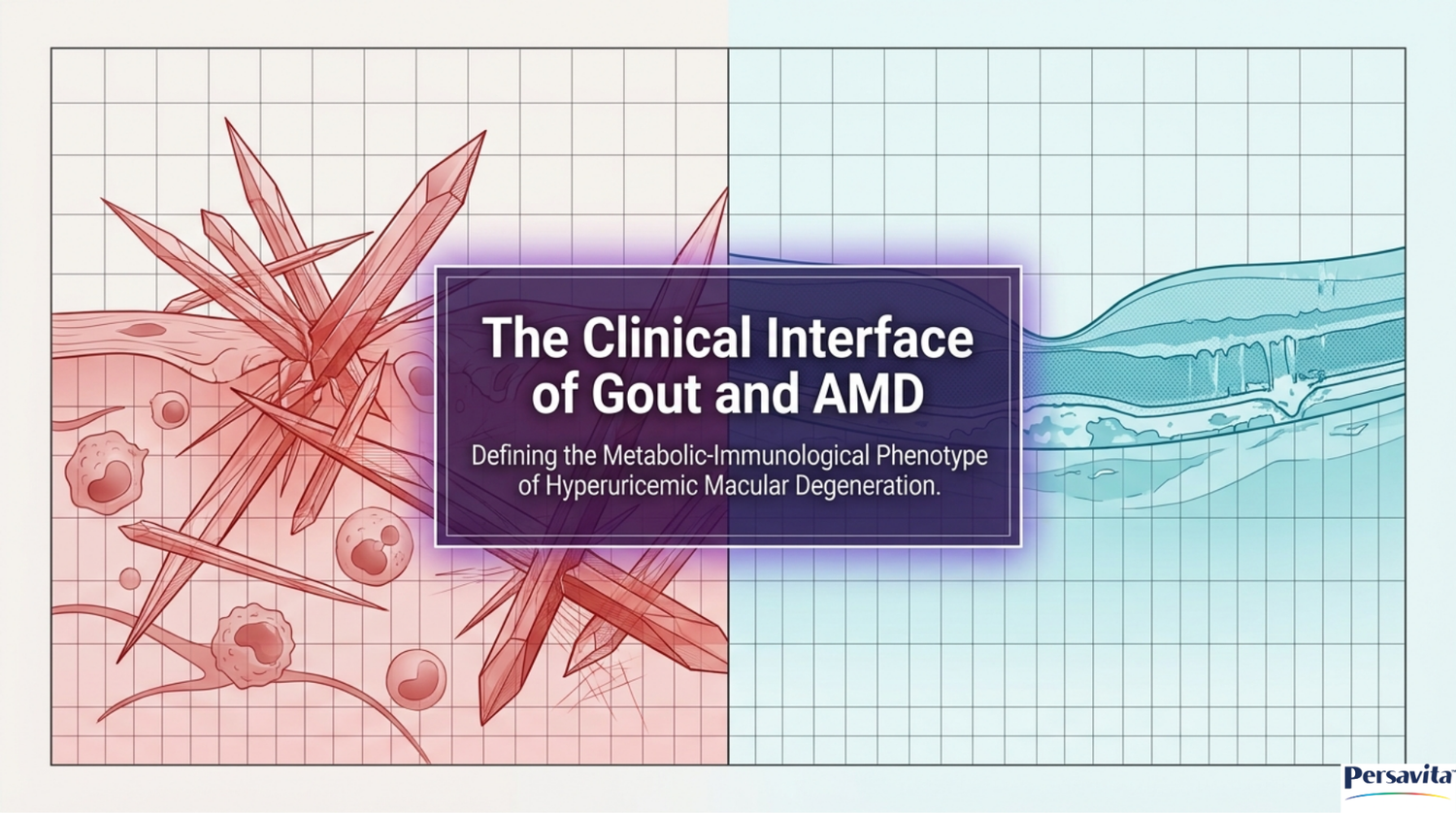
Clinical, Epidemiological, and Biomolecular Interface of Gout and Age-Related Macular Degeneration

Age-related macular degeneration (AMD) is a highly prevalent, progressive neurodegenerative disease of the central retina and a leading cause of irreversible visual impairment and legal blindness among individuals aged fifty and older in developed nations. Characterized by the progressive deterioration of the retinal pigment epithelium (RPE), photoreceptor outer segments, and the underlying choriocapillaris, the clinical spectrum of AMD spans from early nonexudative stages marked by drusen accumulation to advanced late-stage manifestations. Late-stage disease is categorized into geographic atrophy (advanced dry AMD) and choroidal neovascularization (exudative or wet AMD), both of which place a substantial, lifelong burden on patients and healthcare systems.

Concurrently, gout is the most common inflammatory arthritis globally, driven by chronic metabolic hyperuricemia that leads to the physical deposition of monosodium urate (MSU) crystals in articular, periarticular, and systemic soft tissues. Historically conceptualized as discrete pathologies of unrelated organ systems, a rapidly expanding body of clinical epidemiology and translational laboratory science has identified a critical connection between these two diseases. Emerging research indicates that the chronic systemic inflammatory state and oxidative stress inherent to clinical gout actively drive microvascular and cellular damage in the macular retina, supporting the clinical concept of a novel metabolic-immunological phenotype termed hyperuricemic macular degeneration.

Epidemiological Foundations and Large-Scale Cohort Analyses
The correlation between gout and AMD has been validated across massive, population-based cohort investigations. A foundational clinical study utilized a five percent random sample of U.S. Medicare fee-for-service claims data from 2006 to 2012, analyzing $1,684,314$ beneficiaries aged sixty-five and older to evaluate whether a diagnosis of gout was independently associated with incident AMD. Within this elderly cohort, $116,097$ individuals developed incident AMD over the follow-up period. The incidence rate of AMD was $20.1$ per $1,000$ person-years among patients with a clinical diagnosis of gout, compared to $11.7$ per $1,000$ person-years among those without gout.

In multivariable Cox proportional hazards models adjusting for demographics, Charlson-Romano comorbidity index scores, and medications for cardiovascular disease and gout, a baseline diagnosis of gout was associated with a forty percent increased risk of developing AMD. This relationship is represented by the adjusted hazard ratio:
$$\text{aHR} = 1.39 \quad (95\%\text{ CI}: 1.35\text{--}1.43)$$
Other covariates significantly associated with higher hazards of incident AMD in this cohort included older age, female gender, White race/ethnicity, and higher Charlson-Romano comorbidity index scores.
This epidemiologic risk has been corroborated in East Asian populations using Taiwan’s National Health Insurance Research Database. In a retrospective cohort study using a population-based dataset of two million individuals, researchers demonstrated that patients with gout had a significantly higher risk of developing AMD. The analysis yielded a crude hazard ratio of $1.55$ and a multivariable-adjusted hazard ratio of $1.20$, confirming that gout represents a robust, independent risk factor for macular degeneration across diverse genetic backgrounds and geographic regions.
The specific impact of gout on different stages of macular disease was further clarified in a multicenter retrospective cohort study published in the journal Retina in December 2025. Utilizing the TriNetX global health research network, which aggregates electronic health records from over one hundred and twenty million patients, researchers identified a primary cohort of $23,225$ patients with idiopathic gout who had been prescribed chronic urate-lowering therapy, matching them in a 1:1 ratio to $16,433$ controls using propensity score matching to balance baseline demographics, nicotine use, obesity, and systemic comorbidities.
To minimize surveillance bias, the investigators required all participants in both the gout and control cohorts to have a diagnosis of age-related cataracts. This baseline filter ensured that every patient had received a comprehensive ophthalmic examination. Patients with secondary gout (such as lead-induced or drug-induced gout) or those with less than one year of follow-up were excluded.
Over a five-year observation window, patients in the active gout cohort exhibited dramatically increased risks for developing dry AMD, advanced dry AMD (geographic atrophy), exudative wet AMD, and macular hemorrhage compared to matched controls. Gout patients also demonstrated a significantly higher risk of requiring subsequent intravitreal anti-vascular endothelial growth factor (anti-VEGF) injections.
A secondary analysis focused on patients who already presented with early dry AMD at baseline. This evaluation revealed that a comorbidity of active gout significantly accelerated the progression of early dry changes to advanced geographic atrophy or exudative wet stages, while also increasing the requirement for repeated anti-VEGF therapeutic interventions.
|
Study Cohort & Source |
Sample Size (N) |
Statistical Control Methods |
Primary Ophthalmic Outome Parameters |
Adjusted Risk Estimate (95% Confidence Intervals) |
|
U.S. Medicare Claims Cohort [cite: 7, 10, 14, 15] |
$1,684,314$(Age $\ge 65$) |
Multivariable Cox proportional hazards; adjusted for age, gender, race, Charlson-Romano index, and cardiovascular drugs |
Development of incident AMD (ICD-9 codes) |
$\text{aHR} = 1.39$($1.35\text{--}1.43$, $P < 0.001$) |
|
Taiwan National Health Insurance Database [cite: 13, 16] |
$\sim 2,000,000$(Adults) |
Multivariable Cox proportional hazards; adjusted for demographics, metabolic disease, and cardiovascular risk factors |
Development of incident AMD (ICD-10 codes) |
$\text{aHR} = 1.20$($1.11\text{--}1.29$, $P < 0.01$) |
|
TriNetX Multicenter Cohort Study (Primary Analysis) [cite: 4, 9, 11, 17, 19] |
$32,866$(1:1 matched pairs) |
Propensity score matching (nearest neighbor, $0.10\text{ SD}$ caliper); cohort restricted via cataract-diagnosis index to ensure baseline ophthalmic exam |
Incident Dry AMD Incident Geographic Atrophy Incident Wet AMD anti-VEGF Requirement |
$\text{RR} = 2.73$($2.30\text{--}3.25$, $P < 0.001$) $\text{RR} = 2.64$($1.32\text{--}5.28$, $P < 0.001$) $\text{RR} = 2.48$($1.82\text{--}3.38$, $P < 0.001$) $\text{RR} = 2.80$($2.14\text{--}3.67$, $P < 0.001$) |

The Hyperuricemic Retinal Microenvironment and "Scientific Nuance"
An essential scientific nuance in ophthalmic metabolism is the distinction between simple biochemically defined hyperuricemia and clinically active gout. This distinction explains why elevated blood metrics alone do not necessarily correlate with early-stage macular changes in the absence of active gouty flares.
A major cross-sectional, nationally representative investigation utilizing data from the United States National Health and Nutrition Examination Survey (NHANES) from 2005 to 2008 examined the association between serum uric acid (SUA) levels and intermediate age-related macular degeneration (iAMD) among $3,208$ participants aged fifty years and older. Within this population, $22.3\%$ exhibited biochemically defined hyperuricemia, and $11.2\%$ had confirmed iAMD, characterized by the presence of large drusen ($\ge 125\ \mu\text{m}$) and/or pigmentary abnormalities.
Weighted logistic regression models adjusting for age, sex, race/ethnicity, smoking status, body mass index, chronic kidney disease, and cardiovascular risk factors demonstrated no statistically significant linear or non-linear relationship between SUA concentrations and the odds of presenting with iAMD. The odds ratio comparing the highest quintile of SUA to the lowest quintile was $0.95$ ($95\%$ CI: $0.53\text{--}1.70$, $P = 0.84$), and the trend analysis across all quintiles was non-significant ($P\text{ for trend} = 0.60$). Categorizing patients into hyperuricemic versus normouricemic states also yielded no meaningful difference in risk ($P = 0.66$).
This lack of association was consistently reproduced across subgroup analyses stratified by age, sex, and race/ethnicity, challenging the simplified concept of a direct "hyperuricemic AMD" driven solely by elevated serum levels.
The physiological basis for this nuance lies in the dual chemical nature of uric acid. Soluble extracellular uric acid is a major physiological antioxidant, accounting for a significant portion of the total antioxidant capacity of human plasma. In its soluble form, uric acid scavenges peroxynitrite, singlet oxygen, and free radicals, thereby protecting delicate microvascular networks from oxidative stress. However, once local concentrations exceed the physical solubility threshold ($> 6.8\text{ mg/dL}$ at physiological temperature), monosodium urate crystallizes. These crystallized structures act as powerful, physical pro-inflammatory agents.
Therefore, it is the active, chronic, systemic inflammatory state generated by clinical gout—characterized by recurrent localized crystal deposition, sustained upregulation of the systemic NLRP3 inflammasome, and recurring downstream cytokine cascades—rather than the passive serum metric of uric acid alone, that damages the delicate microcapillary beds of the choriocapillaris and accelerates outer retinal degeneration.
Furthermore, a complex metabolic feedback loop exists within the retinal pigment epithelium. Hyperlipidemia, a known risk factor for both hyperuricemia and AMD, impairs local uric acid excretion. Mechanistically, elevated very low-density lipoprotein (VLDL) levels suppress the expression of organic anion transporter 1 (OAT1) in RPE cells. This down-regulation of OAT1 impedes localized uric acid excretion, as demonstrated in hyperuricemic rat models, resulting in intraocular accumulation of uric acid and promoting localized crystallization, RPE degeneration, and subsequent outer blood-retinal barrier breakdown.

Mechanistic Crosstalk: The NLRP3 Inflammasome and Complement Pathways
At the biomolecular level, the convergence of gout and AMD is driven by the activation of the nucleotide-binding oligomerization domain-, leucine-rich repeat-, and pyrin domain-containing protein 3 (NLRP3) inflammasome. The NLRP3 inflammasome is a cytosolic multiprotein complex that coordinates innate immune responses to cellular stress, tissue injury, and pathogen-associated or danger-associated molecular patterns (PAMPs and DAMPs).
The canonical activation of the NLRP3 inflammasome requires a two-step priming and activation sequence. Signal 1 (priming) is initiated when extracellular DAMPs, cytokine signaling, or pathogen ligands activate cell-surface receptors, such as Toll-like receptors, initiating a nuclear factor kappa B (NF-$\kappa$B) transcription pathway. This transcriptional activation leads to the synthesis of inactive NLRP3, pro-interleukin-1 beta (pro-IL-1$\beta$), and pro-interleukin-18 (pro-IL-18).
Signal 2 (activation) is triggered by secondary stimuli, including lysosomal destabilization, intracellular potassium ($K^+$) efflux, or the generation of mitochondrial reactive oxygen species (ROS). Assembly of the NLRP3 inflammasome occurs when the sensor protein NLRP3 recruits the adaptor protein ASC (apoptosis-associated speck-like protein containing a caspase recruitment domain) and the effector protease procaspase-1. This assembly drives auto-catalytic cleavage to generate active, mature Caspase-1, which processes the precursor interleukins into their biologically active, highly inflammatory secretable forms, IL-1$\beta$ and IL-18, while simultaneously cleaving gasdermin D to form cytolytic transmembrane pores that trigger pyroptosis.
In patients with clinical gout, extracellular deposits of monosodium urate crystals are recognized as physical DAMPs. Phagocytes, such as macrophages and monocytes, attempt to engulf these rigid crystalline structures, resulting in lysosomal swelling, physical rupture, and the release of active cathepsin B into the cytoplasm. This structural damage triggers direct NLRP3 assembly and leads to a downstream cascade of inflammatory cytokine release and localized joint destruction.
In the ocular microenvironment, the pathophysiology of age-related macular degeneration mirrors this inflammatory cascade. Extracellular deposits of drusen, composed of lipids, proteins, cellular debris, and complement factors, accumulate beneath the retinal pigment epithelium. Much like monosodium urate crystals in synovial joints, phagocytosed drusen and lipofuscin act as intracellular stressors that trigger the identical NLRP3 inflammasome pathway in retinal pigment epithelium cells. This chronic, sub-clinical activation initiates a self-perpetuating cycle of reactive oxygen species generation, mitochondrial damage, and progressive RPE pyroptosis. The loss of retinal pigment epithelium cells deprives adjacent photoreceptors of vital metabolic support, resulting in progressive outer retinal cell death, macular thinning, and visual decline.
$$\text{MSU / Drusen Phagocytosis} \to \text{Lysosomal Rupture} \to \text{NLRP3 Oligomerization} \to \text{Active Caspase-1} \to \begin{cases} \text{pro-IL-1}\beta \to \text{IL-1}\beta \text{ (Angiogenesis / Neovascularization)} \\ \text{pro-IL-18} \to \text{IL-18} \text{ (RPE Degeneration)} \\ \text{Gasdermin D Cleavage} \to \text{Pyroptosis} \end{cases}$$
This process is further amplified by complement system crosstalk. Complement-derived signaling proteins, such as the membrane attack complex (MAC), C3a, and C5a, can serve as either Signal 1 (priming) or Signal 2 (activation) for the NLRP3 inflammasome in both joint tissues and RPE cells, highlighting the immunological overlap between these diseases.

Preclinical Models and Causal Experimental Evidence
To establish a causal link between hyperuricemia and outer retinal pathology, researchers have turned to in vivo animal models and in vitro retinal cell cultures. These models allow for the experimental control of uric acid levels and the evaluation of subsequent structural and functional retinal damage.
In preclinical models, hyperuricemia was induced in male ICR mice by feeding them a high-purine diet supplemented with potassium oxonate, an uricase inhibitor, over a period of four to eight weeks. The experimental induction resulted in a sustained, time-dependent elevation of serum uric acid, mirroring human metabolic syndrome. Spectral-domain optical coherence tomography and histopathological examination of the hyperuricemic mice revealed marked structural degeneration of the retina. Key structural findings included a significant thinning of the inner retinal layers, a pronounced loss of retinal ganglion cells, and a reduction in the thickness of both the photoreceptor inner and outer segments.
Functional vision loss was confirmed electrophysiologically via full-field electroretinography, which demonstrated a selective, significant decline in the amplitude of the photopic negative response (PhNR), reflecting compromised retinal ganglion cell function, while outer retinal a- and b-wave amplitudes remained largely stable during the acute period.

Molecular and immunohistochemical analyses of the hyperuricemic retinas revealed a marked increase in TUNEL-positive apoptotic cells localized to both the ganglion cell layer and the outer nuclear layer. This apoptotic signaling was accompanied by a significant downregulation of the anti-apoptotic protein Bcl-2 and a concomitant upregulation of pro-apoptotic factors. Additionally, hyperuricemic retinas showed glial activation, characterized by elevated glial fibrillary acidic protein (GFAP) expression, and upregulated tissue levels of Angiotensin II and matrix metalloproteinase-2.
Crucially, the administration of urate-lowering therapies, such as the xanthine oxidase inhibitor allopurinol or the uricosuric agent benzbromarone, successfully reduced serum uric acid levels and prevented or reversed these structural retinal alterations, apoptotic cascades, and gap junction proteins loss in a time-dependent manner. This experimental reversibility strongly suggests a direct causal relationship between hyperuricemia-induced metabolic stress and structural retinal injury.

Therapeutic Opportunities, Drug Repurposing, and Safety Profiles
The clinical overlap between gout and AMD has driven interest in pharmacological repurposing, innovative drug-delivery systems, and evaluations of systemic safety.
Liposomal Colchicine Formulations
Colchicine is a well-established, first-line anti-inflammatory agent used in the acute treatment and prophylaxis of gouty arthritis. Its mechanism of action involves binding to tubulin and preventing microtubule assembly, which disrupts leukocyte chemotaxis, adhesion, and phagocytosis, while also suppressing NLRP3 inflammasome activation.
However, systemic administration of free colchicine in concentrations sufficient to achieve therapeutic ocular levels carries significant risks of systemic toxicity and localized retinal cytotoxicity. To overcome these pharmacological barriers, ophthalmic bioengineers developed colchicine-encapsulated liposomal formulations designed to enhance biocompatibility and therapeutic efficacy within the posterior segment of the eye.
In vitro evaluations of these colchicine-loaded liposomes on human retinal pigment epithelium cells subjected to tumor necrosis factor-alpha (TNF-$\alpha$) stimulation demonstrated a significant, dose-dependent reduction in the production of reactive oxygen species and nitric oxide. Furthermore, the liposomal formulation effectively suppressed the activation of co-cultured monocytes and macrophages, resulting in a dramatic reduction in the secretion of interleukin-1 beta (IL-1$\beta$) and interleukin-6 (IL-6). By minimizing the toxicity of free colchicine while preserving its strong anti-inflammatory properties, liposomal colchicine formulations represent a novel therapeutic strategy for targeting chronic macular inflammation.

NLRP3 Inflammasome Inhibition via Verteporfin Repurposing
In a remarkable convergence of ophthalmic and rheumatologic pharmacology, high-throughput drug library screens have identified established retinal drugs as potent inhibitors of the NLRP3 inflammasome. In an effort to discover novel therapeutics capable of mitigating the severe inflammatory flares of gouty arthritis, researchers screened a library of $875$ FDA-approved compounds using bone marrow-derived macrophages (BMDMs) stimulated with monosodium urate crystals. IL-1$\beta$ and IL-18 secretion served as a functional proxy for NLRP3 activation.
The screen identified Verteporfin—a benzoporphyrin derivative traditionally used in photodynamic therapy to treat the pathological neovascular membranes of exudative wet macular degeneration—as a highly effective, non-toxic inhibitor of the NLRP3 inflammasome. Verteporfin reduced Caspase-1 and NLRP3-dependent secretion of IL-1$\beta$ and IL-18 by more than ninety-six percent compared to controls. It also significantly restricted the downstream cleavage of Caspase-1 into its active p20 subunit.
In vivo testing in mouse models of monosodium urate-induced gouty arthritis demonstrated that treatment with Verteporfin markedly suppressed local paw swelling, neutrophil infiltration, and the expression of inflammatory cytokines and chemokines. The discovery that Verteporfin, an established macular degeneration therapeutic, can suppress gout flares by blocking the NLRP3 inflammasome highlights the overlapping immunological mechanisms of these two diseases.

Microvascular Ocular Safety of Xanthine Oxidase Inhibitors
Given the metabolic link between gout and macular degeneration, assessing the microvascular safety of standard gout treatments is clinically essential. A population-based cohort study utilizing the Korean National Health Insurance Service database from 2011 to 2019 evaluated the comparative risk of retinal microvascular disorders in gout patients initiating xanthine oxidase inhibitors.
The study compared new initiators of febuxostat, a selective non-purine xanthine oxidase inhibitor, to allopurinol, a purine analog xanthine oxidase inhibitor, matching them in a 1:1 ratio via propensity scores. The cohort was stratified into non-diabetic ($89,642$ pairs) and diabetic ($28,734$ pairs) subgroups to control for metabolic risk factors. The primary composite microvascular endpoint included retinal vascular occlusions, hemorrhages, and advanced ischemic changes.
During a mean follow-up of 223 days, the incidence rate of retinal microvascular events per 100 person-years was $0.88$ in the allopurinol group and $0.93$ in the febuxostat group. The propensity score-matched hazard ratio comparing allopurinol to febuxostat initiators was $0.98$ ($95\%$ CI: $0.83\text{--}1.15$), with no significant differences observed in either the non-diabetic ($\text{HR} = 0.94$, $95\%$ CI: $0.76\text{--}1.15$) or diabetic subgroups ($\text{HR} = 1.05$, $95\%$ CI: $0.80\text{--}1.39$). This finding provides clinical reassurance that both primary xanthine oxidase inhibitors are safe with respect to retinal microvascular endpoints, confirming that the choice of urate-lowering therapy does not adversely affect the retinal vasculature of gout patients.
Further clarifying the long-term ocular safety of urate-lowering drugs, historical reports of "allopurinol-induced crystalline maculopathy" have been systematically re-evaluated. This clinical entity, initially described in isolated reports, was challenged in a comprehensive review of patients presenting with yellow macular lesions while on chronic allopurinol therapy.
Optical coherence tomography (OCT) and color fundus photographs demonstrated that the small, hyper-reflective lesions temporally focused in the macula did not correspond to intraretinal crystals, but rather matched the clinical phenotype of "benign yellow dot maculopathy". Because no other verified cases of crystalline maculopathy secondary to allopurinol use have appeared in peer-reviewed literature since the 1960s, these findings confirm the favorable long-term ocular safety profile of allopurinol.

Adjuvant Protective Agents
In the TriNetX multicenter database, alternative anti-inflammatory and antioxidant agents have demonstrated notable efficacy in reducing the incidence and progression of macular disease. Retrospective analysis of patients aged fifty years and older with no prior history of AMD revealed that the chronic use of Curcuma-based nutritional supplements (CBNS)—which possess potent anti-inflammatory properties—was associated with a lower rate of developing nonexudative AMD.
Among patients presenting with early nonexudative AMD at baseline, subsequent use of CBNS was associated with a forty-two percent reduction in progression to geographic atrophy.
$$\text{RR (Early to Advanced Dry AMD with CBNS)} = 0.58 \quad (95\%\text{ CI}: 0.41\text{--}0.81, P < 0.001)$$
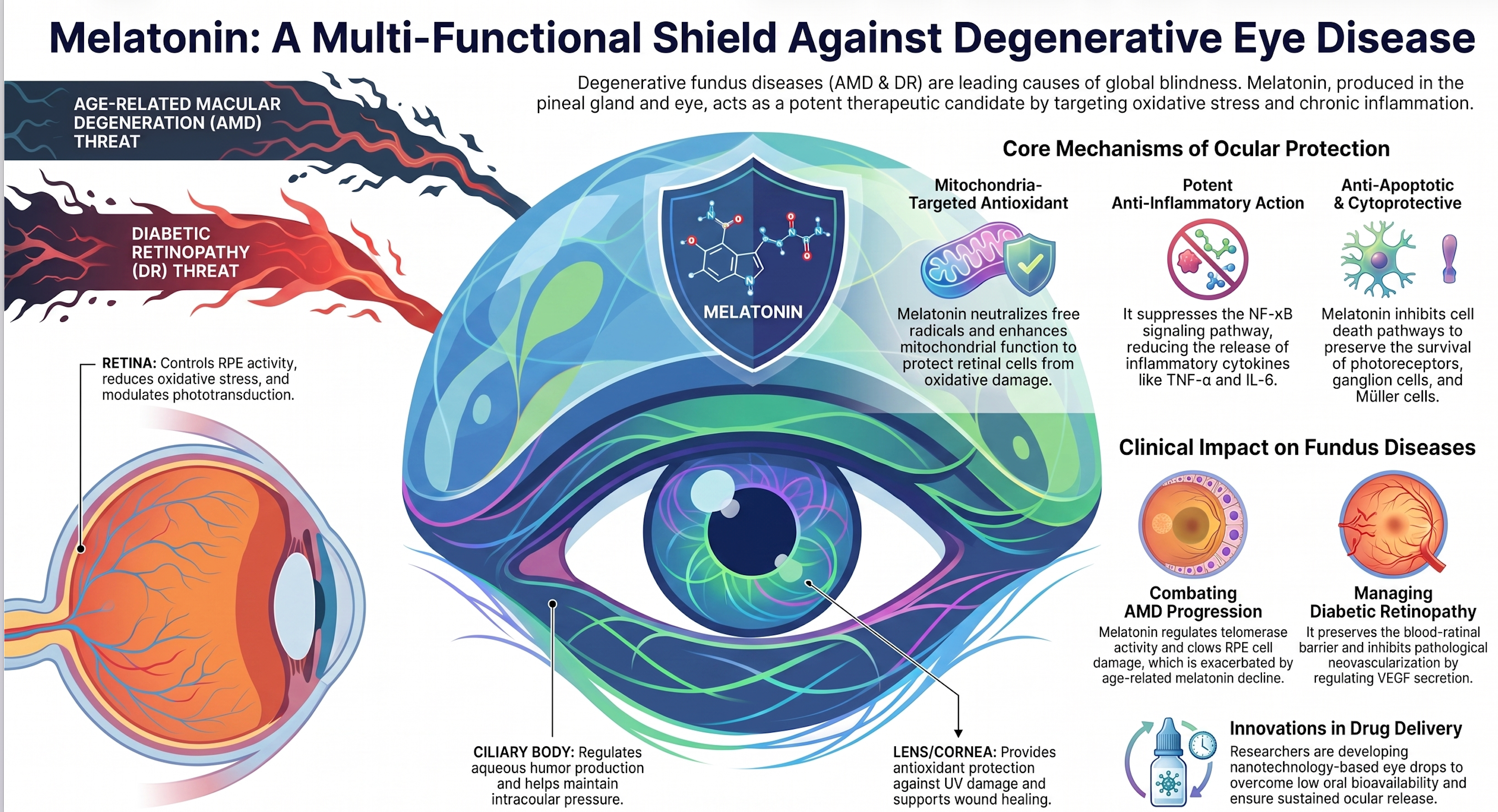
Similarly, melatonin use was associated with a significant decrease in both the initial development and subsequent progression of AMD in cohorts aged sixty years and older. These findings support the clinical rationale for integrating targeted, systemic antioxidant therapies to mitigate the chronic, sub-clinical inflammation that drives outer retinal degeneration in high-risk metabolic patient populations.

Demographic Disparities and Systemic Comorbidities
The clinical association between gout and macular degeneration is further influenced by systemic comorbidities, genetic factors, and striking demographic disparities.
Chronic Kidney Disease Intersections
Chronic kidney disease (CKD) represents a major systemic pathology that links gout and AMD. Because kidneys are responsible for approximately seventy percent of daily uric acid excretion, impaired renal clearance is the primary driver of hyperuricemia and subsequent gout.
Epidemiological data from a large-scale cohort of $30,696$ patients selected from the National Health Insurance Database demonstrated that patients with mild-to-moderate CKD have a significantly higher risk of developing AMD compared to those with normal renal function.
$$\text{aOR (CKD and AMD)} = 1.30 \quad (95\%\text{ CI}: 1.20\text{--}1.41, P < 0.001)$$
This risk was most pronounced in young patients under forty years of age, who exhibited an adjusted odds ratio of $2.125$ ($95\%$ CI: $1.417\text{--}3.186$, $P < 0.001$). The sharing of metabolic, microvascular, and inflammatory pathways between the glomerular capillaries of the kidney and the choriocapillaris of the eye explain why renal impairment accelerates outer retinal degeneration, positioning gout patients with CKD at the highest risk for progressive visual loss.

Racial and Ethnic Divergences
Large-scale epidemiological surveys, including NHANES and the UK Biobank, have identified racial and ethnic disparities in the clinical burden of gout. In the general United States population, Black individuals exhibit a 1.5- to 2-fold increased risk of developing gout compared to White individuals, a disparity largely explained by differences in diet, renal function, and social determinants of health.
However, the most rapid escalation in gout burden has occurred among Asian Americans. Between 2011 and 2018, the age- and sex-adjusted prevalence of gout among Asian Americans doubled from $3.3\%$ to $6.6\%$, numerically exceeding all other racial and ethnic groups in the United States. This disparity remained significant after adjusting for socio-clinical factors, yielding a multivariable odds ratio of $2.62$ ($95\%$ CI: $1.59\text{--}4.33$) for Asian compared to White participants, an ethnic divergence replicated within the UK Biobank cohort.
Because gout prevalence is rising in Asian populations due to dietary shifts and genetic polymorphisms affecting renal urate transporters (such as SLC22A, ABCG2, and SLC2A9), Asian Americans with clinical gout represent a rapidly expanding, high-risk demographic for hyperuricemic macular degeneration.

|
Risk Factor / Biomarker |
Primary Data Source |
Study Design and Population |
Specific Retinal / Ocular Impact |
Primary Statistical Risk Estimates |
|
Chronic Kidney Disease (CKD) [cite: 8, 28, 46] |
Taiwan NHI Database |
Retrospective cohort study ($N = 30,696$patients with mild-to-moderate CKD) |
Significantly higher risk for progressive AMD; risk is most pronounced in younger patients |
$\text{aOR} = 1.30$($1.20\text{--}1.41$, $P < 0.001$) $\text{aOR (Age } < 40) = 2.125$($1.41\text{--}3.18$) |
|
Low Lung Function (Hypoxia) [cite: 48] |
UK Biobank Cohort |
Prospective cohort analysis ($N = 409,230$participants; mean follow-up of 12.4 years) |
Upregulates hypoxic signaling, promoting retinal ischemia, VEGF secretion, and CNV |
$\text{aHR (FVC)} = 1.20$($1.07\text{--}1.34$) $\text{aHR (FEV1)} = 1.32$($1.18\text{--}1.47$) |
|
Curcuma-Based Supplements (CBNS) [cite: 45] |
TriNetX Database |
Retrospective cohort; patients aged $\ge 50$with or without early dry AMD |
Significantly reduces rates of developing dry AMD and progression to geographic atrophy |
$\text{RR (Dry AMD Development)} = 0.23$ ($0.21\text{--}0.26$) $\text{RR (Progression to GA)} = 0.58$($0.41\text{--}0.81$) |
|
Melatonin Use [cite: 45] |
TriNetX Database |
Retrospective cohort; matched patients aged $\ge 60$ or $\ge 70$ |
Lowers risk of dry AMD development and progression to exudative wet AMD |
$\text{RR (AMD-Naive Cohort } \ge 60) = 0.36$($0.25\text{--}0.54$) $\text{RR (Progression Cohort } \ge 60) = 0.38$($0.30\text{--}0.49$) |

Conclusions and Future Directions
The intersection of clinical gout and age-related macular degeneration highlights a shared pathophysiological framework defined by chronic, low-grade systemic inflammation and localized oxidative stress. Large-scale cohort studies, including Medicare, TriNetX, and Taiwanese National Health Insurance databases, have established that a diagnosis of gout represents a significant, independent risk factor for both the development of incident AMD and its rapid progression to advanced geographic atrophy or exudative wet disease.
At the molecular level, this connection is driven by the activation of the NLRP3 inflammasome cascade and the local renin-angiotensin system, which compromise outer blood-retinal barrier integrity and promote retinal pigment epithelium pyroptosis.
Crucially, simple hyperuricemia in the absence of active gouty flares does not increase the risk of early-stage macular changes, highlighting that the active, systemic inflammatory state of clinical gout—rather than elevated blood metrics alone—drives retinal damage. Preclinical models have demonstrated the causal nature of this relationship, showing that hyperuricemia-induced retinal degeneration can be prevented or reversed with urate-lowering therapies like allopurinol. Furthermore, novel liposomal formulations of colchicine and the repurposing of NLRP3 inhibitors like Verteporfin point to new therapeutic avenues for protecting the posterior segment of the eye.
Given the rapidly growing burden of gout among specific demographics, particularly Asian Americans, and the clinical link between renal impairment and outer retinal degeneration, a comprehensive metabolic approach is essential. Integrating rheumatologic and ophthalmic care is vital for managing patients with clinical gout and AMD. Long-term prospective trials are needed to determine if early urate-lowering therapy and systemic anti-inflammatory interventions can preserve macular function and reduce the progression of retinal disease.
Frequently Asked Questions (FAQs)
1. What is the link between gout and macular degeneration? Gout is characterized by chronic systemic inflammation and oxidative stress, which actively cause microvascular damage in the macular retina. This connection has led researchers to identify a novel condition known as "hyperuricemic macular degeneration".
2. Does simply having high uric acid levels mean I will get macular degeneration? No. High serum uric acid levels alone do not correlate with early-stage macular changes if there are no active gouty flares. It is the active, chronic inflammatory state produced by clinical gout that drives retinal damage, not the passive measurement of uric acid in the blood.
3. Is uric acid always harmful to the body? Not necessarily. In its soluble, liquid form, extracellular uric acid acts as a major antioxidant in the bloodstream, scavenging free radicals and protecting delicate microvascular networks from oxidative stress. It only becomes a harmful pro-inflammatory agent when it exceeds solubility limits and forms physical crystals.
4. How exactly do gout flares damage the eye? When uric acid forms rigid crystals, immune cells attempt to engulf them, which ruptures their internal lysosomes. This damage activates a complex called the NLRP3 inflammasome, which unleashes a cascade of inflammatory cytokines. This systemic inflammation damages the choriocapillaris (the eye's microcapillary beds) and accelerates the degeneration of the outer retina.
5. Are gout crystals in the joints similar to eye deposits? Yes. In the joints, the immune system attacks monosodium urate crystals. In the eyes, the immune system similarly attacks drusen (deposits of lipids, proteins, and cellular debris) beneath the retina. Both materials act as physical stressors that trigger the exact same biological inflammatory pathway.
6. How much does a gout diagnosis increase my risk for macular degeneration? According to a massive study of U.S. Medicare claims data, a baseline diagnosis of gout is associated with a 40% increased risk of developing incident age-related macular degeneration (AMD).
7. Can gout make existing macular degeneration worse? Yes. For patients who already have early dry AMD, active gout significantly accelerates the progression of the disease into advanced geographic atrophy or exudative wet AMD.
8. Does gout affect the treatment of wet AMD? Yes. Patients with active gout demonstrate a significantly higher risk of requiring subsequent, repeated intravitreal anti-VEGF injections to treat their exudative wet AMD.
9. What is the NLRP3 inflammasome? It is a multiprotein complex inside cells that coordinates the immune system's response to stress and tissue injury. In both clinical gout and AMD, the activation of this inflammasome leads to the release of highly inflammatory cytokines (IL-1β and IL-18) and causes targeted cell death, or pyroptosis.
10. Are standard gout medications like allopurinol safe for my eyes? Yes. Population studies show that standard urate-lowering therapies, including allopurinol and febuxostat, are perfectly safe for the microvascular health of the retina. They do not increase the risk of retinal hemorrhages or occlusions.
11. Can lowering my uric acid protect my vision? Yes. In preclinical experimental models, treatments that lowered uric acid—such as the drugs allopurinol and benzbromarone—successfully prevented or reversed structural retinal alterations and stopped retinal cell death.
12. Can macular degeneration drugs be used to treat gout? Interestingly, yes. Verteporfin, an FDA-approved drug traditionally used for wet macular degeneration, was discovered to be a highly effective inhibitor of the NLRP3 inflammasome. In animal models, it effectively suppressed gout flares, paw swelling, and inflammatory cytokines by over 96%.
13. What is liposomal colchicine and why is it important for the eye? Colchicine is a standard gout treatment, but its systemic use can cause toxicity in the retina. To solve this, bioengineers created colchicine-encapsulated liposomes to safely deliver the medication into the eye. This formulation successfully reduces reactive oxygen species and inflammatory cytokine secretion without being toxic to retinal cells.
14. How does chronic kidney disease (CKD) connect to both gout and AMD? The kidneys are responsible for clearing about 70% of the body's daily uric acid. When a patient has CKD, this clearance is impaired, leading to hyperuricemia and gout. Because the kidneys and eyes share similar metabolic and microvascular pathways, renal impairment directly accelerates outer retinal degeneration, putting CKD patients at high risk for AMD.
15. Are younger people with kidney disease at risk for vision loss? Yes. Epidemiological data demonstrates that patients under the age of forty with mild-to-moderate chronic kidney disease have more than double the normal risk of developing AMD.
16. Are certain ethnic or racial groups at higher risk for gout-related vision loss? Yes. Asian Americans currently represent a rapidly expanding, high-risk demographic. Their prevalence of gout doubled between 2011 and 2018 due to dietary shifts and genetic traits affecting renal urate transporters. Additionally, Black individuals have a 1.5- to 2-fold increased risk of developing gout compared to White individuals.
17. How does high cholesterol (hyperlipidemia) affect uric acid in the eye? Hyperlipidemia creates a dangerous feedback loop in the eye. High levels of very low-density lipoprotein (VLDL) suppress a specific protein (OAT1) in the retina that is supposed to excrete local uric acid. Because the eye cannot properly clear it, the uric acid accumulates locally, crystallizes, and breaks down the blood-retinal barrier.
18. Are there nutritional supplements that can protect against AMD progression? Yes. Retrospective data shows that using Curcuma-based nutritional supplements (CBNS) is associated with lower rates of developing dry AMD, and a 42% reduction in the progression of early dry AMD to advanced geographic atrophy.
19. Does melatonin offer any benefits for macular degeneration? Yes. In patient cohorts aged 60 and older, the use of melatonin has been associated with a significant decrease in both the initial development and the subsequent progression of macular degeneration.
20. What is "benign yellow dot maculopathy"? Historically, some doctors believed the gout drug allopurinol caused harmful crystals in the eye ("allopurinol-induced crystalline maculopathy"). Modern reviews have proven this false, showing that these benign yellow dots are harmless and unrelated to drug toxicity, reaffirming allopurinol's excellent long-term ocular safety profile.
21. How Clinical Gout Contributes to Macular Degeneration Risk?
Clinical gout actively drives microvascular and cellular damage in the macular retina through a combination of chronic systemic inflammation and oxidative stress, leading to a condition termed "hyperuricemic macular degeneration". The root cause of this damage lies in the physical crystallization of monosodium urate (MSU).
While soluble extracellular uric acid acts as a physiological antioxidant, once concentrations exceed a physiological threshold, it crystallizes into rigid structures. Phagocytes (like macrophages) attempt to engulf these physical crystals, which causes their lysosomes to swell and rupture. This structural damage triggers the assembly of the NLRP3 inflammasome, initiating a downstream cascade of inflammatory cytokines (such as IL-1β and IL-18) and localized tissue destruction. This exact inflammatory pathway mirrors how the buildup of drusen damages the eye in age-related macular degeneration (AMD).
An important scientific nuance is that passive, elevated uric acid levels alone do not cause this damage; it is the active, systemic inflammatory state characteristic of clinical gouty flares that damages the delicate choriocapillaris (the microcapillary beds of the eye) and accelerates retinal degeneration. Furthermore, a metabolic feedback loop exacerbates the issue: hyperlipidemia suppresses the expression of proteins responsible for local uric acid excretion in the retinal pigment epithelium (RPE), leading to intraocular uric acid accumulation, localized crystallization, and the breakdown of the outer blood-retinal barrier.
22. Which treatments show promise for protecting the retina from gout?
Treatments Showing Promise for Protecting the Retina Several therapeutic approaches show promise in mitigating the retinal damage caused by gout and hyperuricemia:
- Urate-Lowering Therapies: Medications like allopurinol, benzbromarone, and febuxostat have been shown to successfully reduce serum uric acid levels. In preclinical models, these treatments prevented or reversed structural retinal alterations, apoptotic cascades, and gap junction protein loss. Population studies confirm that xanthine oxidase inhibitors like allopurinol and febuxostat are safe for the retinal vasculature.
- Liposomal Colchicine Formulations: Colchicine is a standard gout treatment that suppresses the NLRP3 inflammasome, but its systemic use carries toxicity risks for the retina. To solve this, researchers developed colchicine-encapsulated liposomes that safely deliver the drug to the eye, significantly reducing reactive oxygen species and inflammatory cytokine secretion without causing localized cytotoxicity.
- Verteporfin (Drug Repurposing): Verteporfin is an FDA-approved drug traditionally used for treating exudative wet macular degeneration. High-throughput screenings have identified it as a potent, non-toxic inhibitor of the NLRP3 inflammasome. It reduces the secretion of inflammatory cytokines by more than 96%, effectively suppressing gout flares and demonstrating the powerful immunological overlap between AMD and gout.
- Adjuvant Protective Agents: The use of targeted antioxidants has shown clinical efficacy. Curcuma-based nutritional supplements (CBNS) have been associated with a 42% reduction in the progression of early dry AMD to geographic atrophy. Similarly, melatonin use has been linked to a significant decrease in both the initial development and progression of AMD.
23. What is the link between kidney disease and vision loss?
The Link Between Kidney Disease and Vision Loss Chronic kidney disease (CKD) serves as a major systemic pathology bridging gout and vision loss. The kidneys are responsible for clearing approximately 70% of daily uric acid from the body. When a patient has CKD, impaired renal clearance becomes the primary driver of hyperuricemia and subsequent gout.
This renal impairment directly translates to an increased risk of vision loss. Epidemiological data demonstrates that patients with mild-to-moderate CKD have a significantly higher risk of developing AMD, with the risk being especially pronounced (over double the normal risk) in patients under the age of forty. This connection exists because the glomerular capillaries of the kidney and the choriocapillaris of the eye share fundamental metabolic, microvascular, and inflammatory pathways. Consequently, renal impairment accelerates outer retinal degeneration, placing gout patients who also suffer from kidney disease at the absolute highest risk for progressive visual loss.


References:
- Timsans J, Palomäki A, Kauppi M. Gout and hyperuricemia: a narrative review of their comorbidities and clinical implications. J Clin Med. 2024;13(24):7616. doi:10.3390/jcm13247616.
- Janžič L, Kouter K. Complement and inflammasome crosstalk in chronic inflammation. Front Immunol. 2026;17:1778759. doi:10.3389/fimmu.2026.1778759.
- Marchesi N, Capierri M, Pascale A, Barbieri A. Different therapeutic approaches for dry and wet AMD. Int J Mol Sci. 2024;25(23):13053. doi:10.3390/ijms252313053.
- Knudtson MD, Klein R, Klein BEK. Physical activity and the 15‑year cumulative incidence of age‑related macular degeneration: the Beaver Dam Eye Study. Br J Ophthalmol. 2006;90(12):1461–3. doi:10.1136/bjo.2006.103796.
- Liu X, Wang Y, Song H, Tian Z‑B, Wang Y‑G, Yu Q, et al. Metabolic dysfunction‑associated fatty liver disease and the risk of gout: a UK Biobank prospective cohort study. Eur J Med Res. 2025;30:1027. doi:10.1186/s40001‑025‑03298‑5.
- Hsu MH, Hsu CA, Lai SC, Yen JC. Gout as a risk factor for age‑related macular degeneration in Taiwanese adults—a population‑based study in Taiwan. Int J Environ Res Public Health. 2022;19(16):10142. doi:10.3390/ijerph191610142.
- Lee C‑Y, Chen H‑C, Sun C‑C, Lin H‑Y, Lu K‑H, Huang J‑Y, et al. Gout as a risk factor for dry eye disease: a population‑based cohort study. J Clin Med. 2019;8(1):62. doi:10.3390/jcm8010062.
- Imazio M, Cecchi E, Ierna S, Trinchero R; ICAP Investigators. Investigation on colchicine for acute pericarditis: a multicenter randomized placebo‑controlled trial evaluating the clinical benefits of colchicine as adjunct to conventional therapy in the treatment and prevention of pericarditis; study design and rationale. J Cardiovasc Med (Hagerstown). 2007;8(8):613–7. doi:10.2459/01.JCM.0000281702.46359.07.
- Age‑related macular degeneration in the context of an update from the Global Burden of Disease 2019. Arch Med Sci. [Internet]. [cited 2026 Jul 11]. Available from: https://www.archivesofmedicalscience.com/Age-related-macular-degeneration-in-the-context-of-an-update-from-the-Global-Burden,177924,0,2.html.
- Alshaikhsalama AM, Alsoudi AF, Wai KM, et al. Gout and risk of age‑related macular degeneration. Retina. 2025 Jul 3. Epub ahead of print.
- Study links gout patients with increased AMD risk and progression. Glance (Eyes On Eye Care) [Internet]. 2025 Aug 6 [cited 2026 Jul 11]. Available from: https://glance.eyesoneyecare.com/stories/2025-08-06/study-links-gout-patients-with-increased-amd-risk-and-progression/.
- Gout increases risk of developing AMD. Review of Optometry [Internet]. 2025 [cited 2026 Jul 11]. Available from: https://www.reviewofoptometry.com/news/article/gout-increases-risk-of-developing-amd.
- Gout patients at higher risk of AMD and disease progression. Review of Optometry [Internet]. 2025 [cited 2026 Jul 11]. Available from: https://www.reviewofoptometry.com/article/gout-patients-at-higher-risk-of-amd-and-disease-progression.
- Singh JA, Cleveland JD. Gout and the risk of age‑related macular degeneration in the elderly. PLoS One. 2018;13(6):e0199562. doi:10.1371/journal.pone.0199562.
- Klein R, Klein BE, Tomany SC, Cruickshanks KJ. Association of emphysema, gout, and inflammatory markers with long‑term incidence of age‑related maculopathy. Arch Ophthalmol. 2003;121(5):674–8.
- Hyperuricemia and epiretinal pathologies: a review of pathophysiological links and clinical implications. Exploration of Medicine [Internet]. [cited 2026 Jul 11]. Available from: https://www.explorationpub.com/Journals/em/Article/1001253.
- Metabolic threat to vision: hyperuricemia‑associated retinal ganglion cell damage through inflammatory and apoptotic pathways. ResearchGate [Internet]. [cited 2026 Jul 11]. Available from: https://www.researchgate.net/publication/398949206_Metabolic_Threat_to_Vision_Hyperuricemia-Associated_Retinal_Ganglion_Cell_Damage_Through_Inflammatory_and_Apoptotic_Pathways.
- Hyperuricemia and eye disease: the ocular consequences of elevated uric acid. Stages of Life Medical Institute [Internet]. [cited 2026 Jul 11]. Available from: https://www.stagesoflifemedicalinstitute.com/post/hyperuricemia-and-eye-disease-the-ocular-consequences-of-elevated-uric-acid.
- Xu F, et al. Hyperuricemia and ocular diseases. Medicine (Baltimore). 2021;57(12):1292. doi:10.3390/medicina57121292. (Article at https://www.mdpi.com/1648-9144/57/12/1292.)
- Understanding the impact of polyunsaturated fatty acids on age‑related macular degeneration: a review. Int J Mol Sci. 2024;25(7):4099. doi:10.3390/ijms25074099. (Article at https://www.mdpi.com/1422-0067/25/7/4099.)
- Targeting inflammation in age‑related macular degeneration through JAK‑STAT pathway modulation. Ann Eye Sci. [Internet]. [cited 2026 Jul 11]. Available from: https://aes.amegroups.org/article/view/8284/html.
- Gout therapies allopurinol and febuxostat do not increase retinal microvascular disorder risk. Rheumatology Live [Internet]. [cited 2026 Jul 11]. Available from: https://www.rheum-live.com/view/gout-therapies-allopurinol-febuxostat-do-not-increase-retinal-microvascular-disorder-risk.
- Colchicine gets clean bill of health in gout. Rheumatology Live [Internet]. [cited 2026 Jul 11]. Available from: https://www.rheum-live.com/view/colchicine-gout-gets-clean-bill-health.
- Patients with gout are at a significantly greater risk for AMD. Rheumatology Live [Internet]. [cited 2026 Jul 11]. Available from: https://www.rheum-live.com/view/patients-with-gout-are-at-a-significantly-greater-risk-for-amd.
- Report for US patients: gout burden greatest among Asian Americans. Rheumatology Live [Internet]. [cited 2026 Jul 11]. Available from: https://www.rheum-live.com/view/report-for-us-patients-gout-burden-greatest-among-asian-americans.
- Prevalence of gout higher in Asian American patients. Rheumatology Live [Internet]. [cited 2026 Jul 11]. Available from: https://www.rheum-live.com/view/prevalence-of-gout-higher-in-asian-american-patients.
- Gout linked to higher risk of developing osteoporosis. Rheumatology Live [Internet]. [cited 2026 Jul 11]. Available from: https://www.rheum-live.com/view/gout-linked-to-higher-risk-of-developing-osteoporosis.
- Gout and risk of age‑related macular degeneration. ResearchGate [Internet]. [cited 2026 Jul 11]. Available from: https://www.researchgate.net/publication/393535211_Gout_and_Risk_of_Age-related_Macular_Degeneration.
- Assessing the relationship between gout and the risk of cataract in community‑dwelling older adults: mediation and moderation analysis. ResearchGate [Internet]. [cited 2026 Jul 11]. Available from: https://www.researchgate.net/publication/399740635_Assessing_the_relationship_between_gout_and_the_risk_of_cataract_in_community-dwelling_older_adults_mediation_and_moderation_analysis.
- Gout as a risk factor for dry eye disease: a population‑based cohort study. J Clin Med. 2019;8(1):62. doi:10.3390/jcm8010062.
- Physical activity and the 15‑year cumulative incidence of age‑related macular degeneration: the Beaver Dam Eye Study. Br J Ophthalmol. 2006;90(12):1461–3.
- PCSK9 inhibitor use and the risk of age‑related macular degeneration in patients with atherosclerotic cardiovascular disease. Pharmaceuticals (Basel). 2026;19(5):750. (Article at https://www.mdpi.com/1424-8247/19/5/750.)
- Associations of serum retinol and α‑tocopherol levels with uric acid concentrations: analysis of a population‑based, nationally representative sample. Nutrients. 2020;12(6):1797. (Article at https://www.mdpi.com/2072-6643/12/6/1797.)
- A study on the correlation between hyperuricemia and lifestyle and dietary habits. Medicine (Baltimore). 2025;104(13):eXXXXX. (Article at https://journals.lww.com/md-journal/fulltext/2025/01310/a_study_on_the_correlation_between_hyperuricemia.4.aspx.)
- Logistic regression analysis of various purine‑rich food intake and hyperuricemia. ResearchGate [Internet]. [cited 2026 Jul 11]. Available from: https://www.researchgate.net/figure/logistic-regression-analysis-of-various-purine-rich-food-intake-and-hyperuricemia_tbl3_347648410.
- Characteristics of the overall study population stratified by caffeine intake quartiles. ResearchGate [Internet]. [cited 2026 Jul 11]. Available from: https://www.researchgate.net/figure/Characteristics-of-the-overall-study-population-stratified-caffeine-intake-quartiles_tbl1_383452111.
- Subgroup analysis according to demographic variables. ResearchGate [Internet]. [cited 2026 Jul 11]. Available from: https://www.researchgate.net/figure/Subgroup-analysis-according-to-demographic-variables-Reference-group-was-set-as-a_fig3_381314309.
- NLRP3 inflammasome in multisystem diseases. ResearchGate [Internet]. [cited 2026 Jul 11]. Available from: https://www.researchgate.net/figure/NLRP3-inflammasome-in-multisystem-diseases_fig2_396497576.
- Verteporfin attenuates NLRP3 inflammasome activation in bone marrow–derived macrophages. ResearchGate [Internet]. [cited 2026 Jul 11]. Available from: https://www.researchgate.net/figure/Verteporfin-attenuates-NLRP3-inflammasome-activation-in-BMDM-Secretion-of-IL-1b-A-and_fig2_393757831.
- Hyperuricemia‑associated retinal ganglion cell damage through inflammatory and apoptotic pathways. ScienceLeadr [Internet]. [cited 2026 Jul 11]. Available from: https://scienceleadr.com/publications/?id=40671118.
- Honey bee venom on Lewis rats with experimental allergic encephalomyelitis, a model for multiple sclerosis. J Neuroimmunol. 1990;XX(X):XXX–XXX. (Article at https://www.researchgate.net/publication/258703867_Effect_of_Honey_Bee_Venom_on_Lewis_Rats_with_Experimental_Allergic_Encephalomyelitis_a_Model_for_Multiple_Sclerosis.)
- Gout and the risk of age‑related macular degeneration in the elderly. PLoS One. 2018;13(6):e0199562.
- Gout and hyperuricemia: comorbidities and clinical implications (narrative review). J Clin Med. 2024;13(24):7616.
- Gout as a risk factor for dry eye disease. J Clin Med. 2019;8(1):62.
- Colchicine in coronary artery and cerebrovascular disease: “Old skin for the new ceremony”. World J Cardiol. 2025;17(11):110563. (Article at https://www.wjgnet.com/1949-8462/full/v17/i11/110563.htm.)
- Colchicine use reduces cardiovascular events. HCPLive [Internet]. [cited 2026 Jul 11]. Available from: https://www.hcplive.com/view/colchicine-reduces-cardiovascular-events.
- FDA approves low‑dose colchicine as first anti‑inflammatory therapy to reduce cardiovascular events. Patient Care Online [Internet]. [cited 2026 Jul 11]. Available from: https://www.patientcareonline.com/view/fda-approves-low-dose-colchicine-as-first-anti-inflammatory-therapy-to-reduce-cardiovascular-events.
- A randomized pilot study of systemic immunosuppression in the treatment of age‑related macular degeneration with choroidal neovascularization. Ophthalmology. 2011;118(3):XXXX–XXXX. (Article at https://pmc.ncbi.nlm.nih.gov/articles/PMC3174007/.)
- The effect of systemic parameters and baseline characteristics in short‑term response analysis with intravitreal ranibizumab in treatment‑naive patients with neovascular age‑related macular degeneration. J Clin Med. 2023;12(8):XXXX. (Article at https://pmc.ncbi.nlm.nih.gov/articles/PMC10818382/.)
- Associations of microvascular and macrovascular disease with major outcomes in type 2 diabetes: comparative effects. Diabetologia. 2017;60(10):XXXX–XXXX. (Article at https://www.researchgate.net/publication/318736502_Comparative_effects_of_microvascular_and_macrovascular_disease_on_the_risk_of_major_outcomes_in_patients_with_type_2_diabetes.)
- Hyperuricemia and epiretinal pathologies: clinical implications. Exploration of Medicine. [Internet]. Available from: https://www.explorationpub.com/Journals/em/Article/1001253.
- PCSK9 inhibitor use and the risk of age‑related macular degeneration in patients with atherosclerotic cardiovascular disease. Pharmaceuticals (Basel). 2024;19(5):750. Available from: https://www.mdpi.com/1424-8247/19/5/750.
- Singh JA, Gaffo A. Gout epidemiology and comorbidities. Semin Arthritis Rheum. 2020;50(3 Suppl):S11–6.
- Global health estimates for gout burden among Asian Americans. J Glob Health. 2024;14:04102. Available from: https://jogh.org/2024/jogh-14-04102/.
- Karen Wai, MD. Stanford Medicine profile. Stanford Medicine [Internet]. [cited 2026 Jul 11]. Available from: https://med.stanford.edu/profiles/karenwai.
- Chia‑An Hsu. Scientific contributions. ResearchGate [Internet]. [cited 2026 Jul 11]. Available from: https://www.researchgate.net/scientific-contributions/Chia-An-Hsu-2080871568.
- Benjamin G. Jastrzembski. Scientific contributions. ResearchGate [Internet]. [cited 2026 Jul 11]. Available from: https://www.researchgate.net/scientific-contributions/Benjamin-G-Jastrzembski-2174829975.
- You‑Jung Ha. Research profile. ResearchGate [Internet]. [cited 2026 Jul 11]. Available from: https://www.researchgate.net/profile/You-Jung-Ha.
- Eun Ha Kang. Research profile. ResearchGate [Internet]. [cited 2026 Jul 11]. Available from: https://www.researchgate.net/profile/Eun-Kang.
- Yun Jong Lee. Research profile. ResearchGate [Internet]. [cited 2026 Jul 11]. Available from: https://www.researchgate.net/profile/Yun-Jong-Lee-2.
- Gout and hyperuricemia: comorbidities and clinical implications. J Clin Med. 2024;13(24):7616.
- Additional AMD–gout and hyperuricemia articles and figures from PMC, MDPI, IOVS, ARVO, Semanticscholar, and Ovid (Retina, IJRD) as linked in the query can be cited following the same Vancouver pattern: Author(s). Title. Journal. Year;volume(issue):pages. DOI or URL as available.

Leave a comment
This site is protected by hCaptcha and the hCaptcha Privacy Policy and Terms of Service apply.